Interview
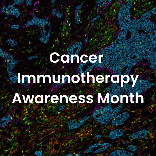
Interview with Dr. Janis Taube: raising awareness about cancer immunotherapy
Posted on:
Immunotherapy is a growing area in cancer research, which aims to harness the body’s immune response against cancerous cells. In fact, immunotherapies are now some of the most promising treatments for cancer available today. But despite their success thus far—and despite ongoing efforts to improve them—there remains much about them that we do not understand: how they work, what determines their success rate in individual patients, and why some patients with the same tumor type respond differently than others under the same therapeutic approach.
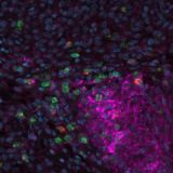
The field of spatial biology is an exciting and fast-moving area. It has the potential to revolutionize immunotherapy by integrating knowledge obtained through multiplex immunofluorescence (mIF) and providing critical biological insights into cancer development and therapy response. Deepening our spatial biology understanding using mIF tools is essential for developing targeted immunotherapies; therefore, improved cancer treatments will result from increased adoption of spatial technologies.
To mark Cancer Immunotherapy Awareness Month, we sat down with Dr. Janis Taube to explore potential ways forward. Janis Taube, The George W. Hambrick, Jr. M.D. and Thomas G. Olsen, M.D. Professor of Dermatology division of Johns Hopkins Hospital in Baltimore, MD. She also serves as a Professor of Dermatology, Professor of Oncology, Professor of Pathology at the Johns Hopkins University School of Medicine, and a Scientific Advisory Board Member at Lunaphore. Dr. Taube is a world-leading expert who developed the PD-L1 immunohistochemical (IHC) assay and scoring system for the first-in-human anti-PD-1 and anti-PD-L1 clinical trials, versions of which are now FDA-approved and mainstays of cancer treatment. Her research primarily focuses on immune evasion by solid tumors—specifically studying the PD-1/PD-L1 axis—and identifying potential biomarkers that predict response to novel immunotherapies.
1. How did you get involved in biomarker research?
There were reports that cancers, including melanoma, expressed PD-L1 as a mechanism of immune evasion. When I first started my post-doctoral fellowship, I was curious to investigate at which phase of melanomagenesis PD-L1 became expressed. We gathered nevi, melanoma in situ, primary and metastatic melanomas to answer this question and developed an IHC assay for PD-L1. We found that PD-L1 expression was not related to the type or stage of melanocytic lesion but to the presence of an immune response. This led us to develop our PD-L1-mediated adaptive immune resistance hypothesis. We then tested this hypothesis by correlating PD-L1 expression within the tumor microenvironment (TME) with response to anti-PD-(L)1-based therapies.
2. The tumor microenvironment (TME) has an essential role in cancer progression. How can the field further harness the TME to accelerate the development of new therapeutic strategies?
A better understanding of components that lead to tumor progression, e.g., myeloid-derived suppressor cells or proteins that lead to immune exclusion in tumors, allows these different components to be targeted in a rational way, facilitating novel combinatorial therapeutic strategies. In addition, if a therapy is shown to be effective in one tumor type, it is possible to characterize the TME of other tumors to see if they show similar features and thus may also be likely to respond to the same regimen.
3. Why is spatial biology the new frontier for advancing our understanding of cancer immunology?
Related Articles
Immuno-oncology: learn from the experts, Prof. Ascierto and Dr. Madonna
Posted on 29 Jun 2022
Read Post