The technology behind COMET™
Our unique approach to spatial biology.
An innovative technology for every lab
Lunaphore is an innovative company that develops cutting-edge technology to accelerate spatial biology adoption through automation, quality, reproducibility, throughput and flexibility.
While our current flexible and open system is mainly designed for fundamental and translational research, our aim is to develop tools that will ultimately pave the way to the clinic.
Here, we present the core technology behind our instrumentation that is at the source of the benefits provided by our laboratory solutions.

A game-changing chip technology
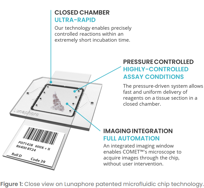
Our core innovation is a unique patented microfluidic chip.
The microfluidic chip is clamped against the tissue section on a standard microscope slide forming a closed reaction chamber (Figure 1).
The delivery of reagents happens through the microfluid channels under highly controlled conditions. Precise reagent flowrates, chamber pressure, reaction temperature and incubation time have been finetuned for optimal performance.
At the end of the protocol, the clamp is released, allowing the reuse of the slide.
Fast Fluidic Exchange (FFeX™) technology
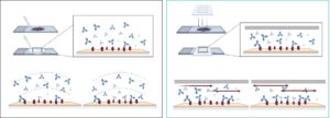
Standard incubation mainly relies on passive diffusion of the antibodies to the targeted epitopes (Figure 2). This results in long incubation times and staining non-uniformity.
Our microfluidic chip performs dynamic incubation through pressured-controlled reagent flow rate and direction. The shallow chamber limits vertical diffusion time, accelerating epitope-reagent exchanges. The combination of the shallow chamber and the active flow provides an almost instantaneous reagents exchange called Fast Fluidic Exchange (FFeX™) (Figure 2).
FFeX™ allows fast, uniform and reproducible staining across the tissue sample, decreasing the incubation time from several hours to only a few minutes.

Figure 2: Principles of standard incubations vs Lunaphore’s FFeX™ incubation.
Benefits of FFeX™
Fast incubation
Thanks to the active flow in the shallow microfluidic chamber, the incubation time is reduced from several hours to only a few minutes.
Precision
The microfluidic chip allows highly dynamic and controlled tuning of assay conditions, including flow rate, flow pressure and chamber temperature.
Staining uniformity
The tightly controlled and almost instantaneous exchange of reagents inside the chip allows a uniform staining of the tissue samples.
Reproducibility
In addition to staining uniformity, assay reproducibility is ensured by the fast incubation that decreases the exposure of tissues to potentially damaging conditions.

Sequential immunofluorescence (seqIF™)
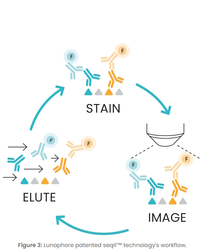
COMET™’s approach to multiplexing is based on the sequential immunofluorescence (seqIF™) technology [1], a process involving successive cycles of staining-imaging-elution (Figure 3), without any human intervention.
During staining, two antigens are detected by conjugation-free primary antibodies and labelled by two secondary antibodies, in addition to counterstaining (DAPI). After staining, images are acquired through the microfluidic chip’s imaging window by COMET™’s integrated microscope. The sequence finishes with the gentle elution of all antibodies that ensures epitope stability and preservation of tissue integrity. Once elution is complete, a subsequent cycle starts, and two new markers are stained and imaged.
All seqIF™ steps are fully automated but parameter tuning is possible as well at every step of seqIF™ cycle.
[1] Rivest, F., Eroglu, D., Pelz, B. et al. Fully automated sequential immunofluorescence (seqIF) for hyperplex spatial proteomics. Sci Rep 13, 16994 (2023).
Available from: https://doi.org/10.1038/s41598-023-43435-w
Benefits of seqIF™
Standard antibodies
The incubation and imaging of only two markers per seqIF™ cycle facilitates hyper-plex assay development through off-the-shelf, non-conjugated antibodies and transfer of existing know-how.
Tissue preservation
The use of a proprietary gentle elution buffer allows to preserve the tissue integrity.
Automation
SeqIF™ on COMET™ is fully automated from sample to ready-to-use data, while preserving the users’ optimization freedom.
Reproducibility
In addition to tissue preservation, the use of non-conjugated antibodies and assay automation, highly reproducible results are generated thanks to controlled conditions.

Unmatched reproducibility
Our seqIF™ technology achieves consistent results across multiple experimental parameters.
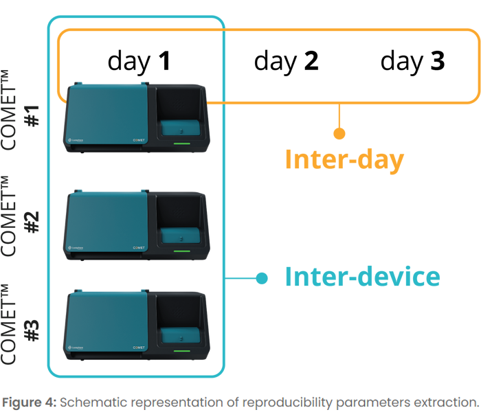
We demonstrated that reproducible staining and imaging can be achieved across different days, multiple devices, and multiple runs in the same device, with less than 15% coefficient of variation of the signal-to-background ratio across all tested parameters.
To access the full quality data or know more about the COMET™ technology, contact us at info@lunaphore.com.