press-releases
Lunaphore announces new data with multiple research collaborators highlighting both the COMET™ platform and LabSat® instrument at the Society for Immunotherapy of Cancer (SITC) 37th Annual Meeting
Date
LAUSANNE, Switzerland – November 10, 2022 – 3:00 pm (CET) – Lunaphore, a Swiss life sciences company developing technology to enable spatial biology in every laboratory, today announced multiple new data sets presented in collaboration with the Geneva University Hospitals, the SIB Swiss Institute of Bioinformatics; the University of Bern, the National Tumor Institute Fondazione G. Pascale in Naples; and Visiopharm. The research will be presented in four posters at the 2022 Society for Immunotherapy of Cancer (SITC) 37th Annual Meeting. The new studies prove the versatility of the expanding applications of Lunaphore’s fully automated, high-throughput platform for hyperplex analysis, COMET™, to map the tumor microenvironment (TME) of lung and colorectal cancer tissues, and the capabilities of LabSat®, the company’s compact and flexible automated tissue staining instrument for chromogenic immunohistochemistry (IHC), multiplex immunofluorescence (mIF) and TSA-based multiplexing.
“The data we are presenting, in partnership with some of the leading research institutions and healthcare technology companies in the world, bring us closer to a future where healthcare professionals can use spatial biology in the clinic to drive treatment decisions,” said Ata Tuna Ciftlik, Ph.D., Chief Executive Officer of Lunaphore. “We have seen this groundbreaking technology applied now to multiple types of cancer, with outstanding efficiency and clear, consistent answers to some of the most difficult problems researchers are trying to solve. We look forward to future collaborations that will further our mission to bring spatial biology and the insights it provides into clinical use”
Mapping the tumor microenvironment with sequential immunofluorescence, an automated image analysis pipeline, and spatial metrics (Mason, D et al.)
The data presented in partnership with Visiopharm, a world leader in AI-driven precision pathology software, outline the synergy created by analyzing the rich sources of data produced by the Lunaphore COMET™ platform with Visiopharm’s dedicated Phenoplex™ workflow for image analysis. Together, the process can extract information from the data sets using AI to deliver single-cell phenotypic information and biodistribution, providing valuable insight into the spatial composition of the TME.
In this study, researchers interrogated tumor composition with the COMET™ platform and Oncotopix® Discovery Phenoplex™, looking at specific cellular phenotypes of interest like proliferating tumor cells, proliferating T cells, immunosuppressive macrophages, and antigen-presenting cells and their interactions, with a focus on the PD-1/PD-L1 pathway. The combination of hyperplex staining with COMET™, advanced image analysis with Oncotopix® Discovery Phenoplex™, and in situ cellular phenotyping allowed researchers to identify tissue composition within the tumor. This workflow demonstrated the potential of analyzing the spatial distribution of specifically phenotyped cells in the TME in order to identify reliable biomarkers that can predict whether certain patients may respond to specific therapies.
Mapping the heterogeneous colorectal microenvironment with multiplexed imaging and machine learning (Cabrera-Gil, B et al.)
In collaboration with the Geneva University Hospitals and the Swiss Institute of Bioinformatics (SIB), researchers looked at the use of multiplex imaging coupled with powerful image analysis tools to highlight important phenotypic differences between patients with microsatellite stable (MSS) and microsatellite instable (MSI) colorectal cancer (CRC). The majority of CRC patients present the MSS CRC type. However, only rare patients from this group respond to immunotherapies currently on the market. Response to immunotherapies is much more frequent in patients with MSI CRC tumors, reflecting potential tumor vulnerability. This emphasizes the importance of understanding the complexities of this disease to identify the patients that are likely to benefit from these treatments and determine if similar treatment options might be available to patients with MSS CRC.
Based on data generated using Lunaphore’s COMET™ platform with a multiplex sequential immunofluorescence (seqIF™) panel of 12 biomarkers, researchers found substantial differences between MSI and MSS CRCs. They were also able to highlight heterogeneity within MSS tumors. Characterizing the TME in this disease state may help define both the prognosis of tumors and the likelihood of response to immune checkpoint therapy. To leverage the advantages of immune modulation in their treatment, the importance of a more precise characterization of the immune response to cancers is essential.
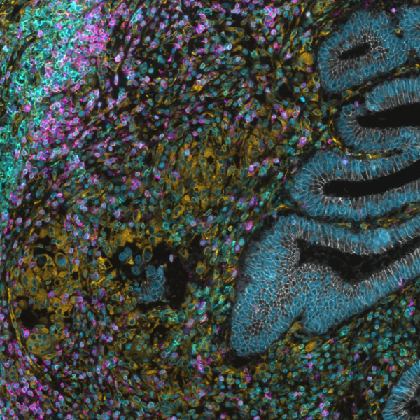
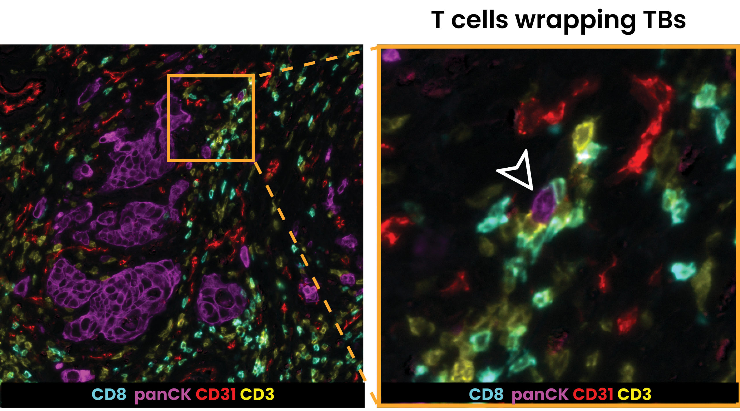
Characterization of tumor budding and the tumor microenvironment in colorectal cancer using hyperplex immunofluorescence (Demir, C et al.)
Research with Prof. Inti Zlobec, Professor of Digital Pathology at the University of Bern, focused on characterizing tumor buds (TBs) in colon cancer as a potential biomarker to improve patient stratification and treatment planning. TBs are recognized as an independent prognostic factor in a variety of solid cancers. In order to test the hypothesis that a subset of TBs represents more aggressive and malignant tumor activity, researchers designed a seqIF™ panel to characterize TBs and the TME.
The 26-plex panel generated on COMET™ allowed for the discrimination of TB signatures within the depth of tumor-stroma interactions and the extraction of valuable TB expression. The analysis will continue with the goal of gathering crucial information about TBs phenotypes and their “cellular neighborhood.” This will finally serve to better identify novel immunograms of these cellular entities, thus better defining their therapeutic potential to advance personalized medicine.
Development of a multiplex test for predicting response to combined immunotherapies in patients with metastatic melanoma (Madonna, G et al.)
Research conducted in collaboration with Dr. Paolo Ascierto, Director of the Unit of Melanoma, Cancer Immunotherapy and Innovative Therapy at the National Tumor Institute Fondazione G. Pascale in Naples highlighted the first steps toward stratification of patients with advanced melanoma who received a combination of immune checkpoint inhibitors (ICIs) in an effort to obtain clinically relevant biomarkers that can predict response to these therapies. In this study, researchers retrospectively analyzed skin metastasis samples from patients with melanoma who were treated with combined ICI, with mixed results seen, using a Tyramide Signal Amplification (TSA) multiplex immunohistochemistry staining for PD-L1, CD8, and LAG-3 expression, followed by DAPI counterstaining, developed and optimized using the Lunaphore LabSat® platform.
This analysis using LabSat® showed that patients who responded to combined ICI therapy showed a statistically significant increase in CD8+ single-positive cell frequency compared to non-responders, and non-responder patients displayed statistically significant increases of PD-L1+ single-positive cell frequency and increased frequency of double CD8+PD-L1+ positive cells, previously found to be a poor prognostic in multiple cancer types. Insights gleaned from this analysis are preliminary evidence of the predictive value of a spatial biomarker test developed using LabSat® to assess whether patients with metastatic melanoma would respond to a certain combination of immunotherapies. A more detailed analysis using larger retrospective and prospective cohorts is ongoing, to further demonstrate clinical relevance.
To learn more about Lunaphore’s presentations and activities at the Society for Immunotherapy of Cancer (SITC) 37th Annual Meeting, please visit: https://lunaphore.com/our-events/sitc-2022/
About Lunaphore
Lunaphore Technologies S.A. is a Swiss company born in 2014 with the vision of enabling spatial biology in every laboratory. Lunaphore has developed a game-changing chip technology that can extract spatial proteomic and genomic data from tumors and transform any simple assay into multiplex spatial biology without complexity. Lunaphore empowers researchers to push the boundaries of research to ultimately develop the next-generation personalized therapies. For further information on Lunaphore and its products, please visit www.lunaphore.com.
About COMET™
COMET™ is a fully automated, sequential immunofluorescence (seqIF™) instrument, able to perform hyperplex staining and imaging, producing high-quality data in a robust and reproducible manner. With superior tissue profiling capabilities, the system allows multiplex analysis of up to 40 different spatial markers per tissue slide without human intervention. COMET™ has a wide range of research applications, allowing for a dramatic improvement in the understanding of disease pathology in areas such as immuno-oncology, neuroscience, and infectious diseases. To learn more about the COMET™ platform, please visit: https://lunaphore.com/products/comet/
About LabSat®
LabSat® is an automated platform designed to perform automated staining of tissue section samples in histological applications. The system’s software interface supports users in creating and executing staining protocols for chromogenic IHC, multiplex immunofluorescence (mIF), and TSA-based multiplexing. The platform enables the generation of high-quality results with excellent reproducibility ensuring tissue preservation. To learn more about the LabSat® platform, please visit: https://lunaphore.com/products/labsat/
For further information contact:
Irene Tamayo
Lunaphore Corporate Communications
Email: [email protected]