Spatial Biology Education

Advancing efficient antibody elution, epitope integrity and tissue preservation on the COMET™ platform

Blog: Advancing efficient antibody elution, epitope integrity and tissue preservation on the COMET™ platform

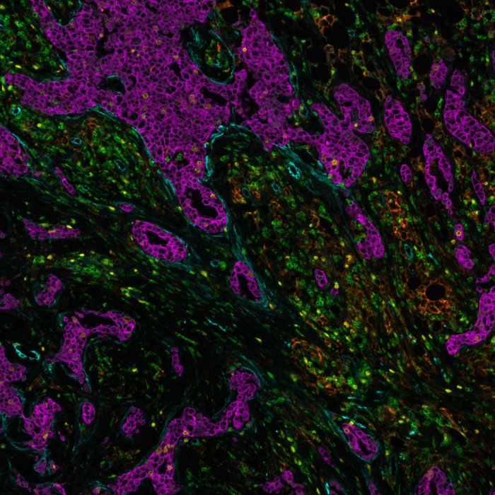
Spatial proteomics is a rapidly evolving field that allows the detection of proteins within biological tissues. The simultaneous detection of protein localization and expression helps scientists and clinicians gain deeper insights into the molecular and cellular interactions within complex tissues. Multiplex immunofluorescence (mIF) is a well-established method in spatial proteomics that enables the detection of multiple protein biomarkers simultaneously on the same tissue section.

Designing a robust and reliable multiplex proteomics experiment requires careful planning to ensure successful detection of multiple protein biomarkers, while maintaining sample integrity. Lunaphore’s Fast Fluidic Exchange (FFeX™) technology is a cutting-edge microfluidics platform that streamlines antibody-based staining, imaging and elution, allowing the efficient detection of protein targets while preserving tissue and epitope stability. The FFeX™ technology enables a rapid immunostaining workflow by drastically reducing incubation times, while maintaining high antigen detection efficiency, resulting in robust and consistent spatial biomarker detection.  
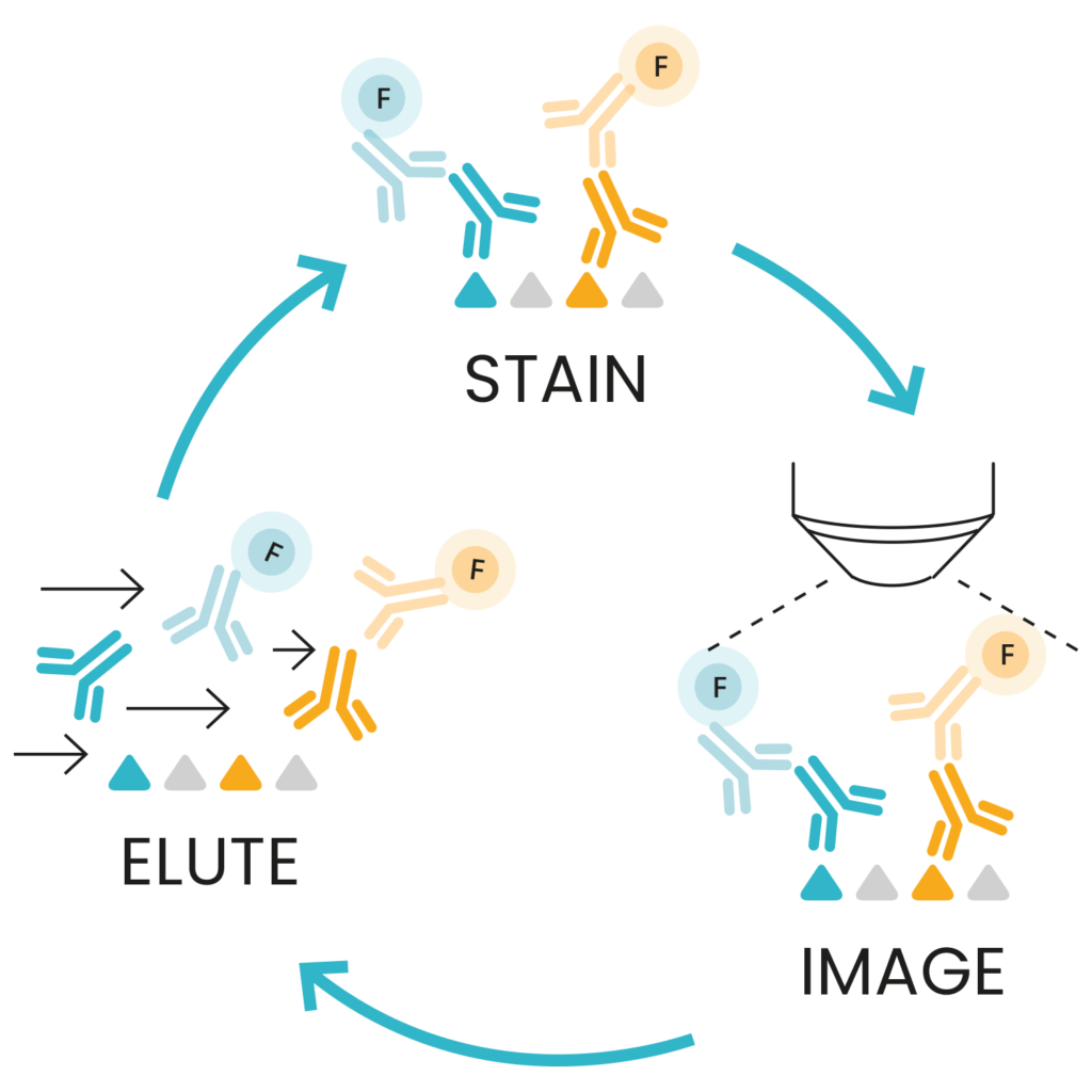
 

The seqIF™ workflow 

Sequential immunofluorescence1 (seqIF™) is an innovative technology with the potential to transform hyperplex spatial proteomics. seqIF™ provides a streamlined alternative to lengthy workflows that require labor-intensive and costly antibody customization through barcoding or conjugation. seqIF™ also addresses reproducibility challenges and tissue damage due to repetitive staining cycles associated with cyclic immunofluorescence methods. 

The seqIF™ workflow is based on repeated sequences of staining, imaging, and antibody elution on the same tissue section as shown in Figure 1. This process utilizes Lunaphore’s FFeX™ technology in use with proprietary reagents and it is designed to maximize the number of detectable targets, while preserving tissue integrity, tissue morphology, and epitope stability, ensuring reliable and consistent results. 

Figure 1: seqIF™ consists of sequential cycles of staining with unconjugated primary antibodies that are then bound by fluorophore-conjugated secondary antibodies, imaging of the tissue followed by gentle elution.


seqIF™ antibody elution efficiency: no compromises on epitope integrity and tissue preservation 

seqIF™ is used to spatially resolve protein biomarkers within tissue sections. Efficient removal of bound antibodies after each staining cycle is critical to avoid signal carry-over or overlap in subsequent imaging cycles. Lunaphore’s fully automated seqIF™ technology uses a gentle elution step that ensures complete removal of antibodies, while preserving the tissue for iterative cycles of staining. A high antibody elution rate is achieved using the FFeX™ microfluidic-based technology that allows fast, uniform, and reproducible elution across the entire tissue sample, ensuring that staining and antibody elution steps are both thorough and non-damaging to the tissue. The antibody elution efficiency on COMET™ has been evaluated across several tissue types and has demonstrated over 95% antibody elution efficiency. Figure 2 shows three examples of staining and post-elution images of the same tissue, and their corresponding antibody elution performance following our COMET™ elution protocol. You can learn more about elution efficiency on COMET™ in this technical note. 

Figure 2: Representative images and quantification to show elution efficiency, tested across more than 15 tissue types. All markers tested have optimal elution efficiency (>95%) on COMET™. Scale bars: 50 µm.

Lunaphore’s gentle elution process enables not only the highly efficient removal of antibodies but also the preservation of epitope stability. Epitope stability is essential for maintaining the integrity of the protein targets throughout multiple staining and elution cycles. The seqIF™ method maintains epitope stability by employing controlled temperature conditions and optimized reagent formulations that preserve epitope integrity. Figure 3 demonstrates that after 20 cycles of seqIF™, the immunoreactivity of epitopes has been maintained, which is essential for accurate and reliable detection of multiple biomarkers from a single tissue sample.

Figure 3: Marker epitope stability over 20 elution cycles on human FFPE tonsil sections. Scale bars: 50 µm.

A recently published study1 highlighted that this approach of mIF can handle up to 40-plex staining panels on FFPE and frozen tissue sections without compromising tissue morphology or causing significant structural damage. Moreover, the same slide can be used to perform any additional downstream characterization, such as H&E or transcriptomics, without the need of tissue transfer steps or additional serial tissue sections. The ability to generate same-section spatial multiomic data is a powerful approach not only for studying morphological changes due to pathological or therapeutic interventions but also to characterize molecular phenotypes at the single-cell level, understand cell distance and dynamics, and measure cell infiltration in a specific tissue niche.  

In conclusion, Lunaphore’s fully automated seqIF™ technology enables spatial hyperplex proteomic studies by ensuring high elution efficiency, maintenance of epitope stability, and preservation of tissue integrity. This innovative approach to mIF provides new opportunities for comprehensive tissue analysis, particularly in fields of immuno-oncology and neuroscience, where understanding the spatial distribution and interaction of multiple biomarkers is essential.  

References: 

  1. Rivest F, Eroglu D, Pelz B, et al. Fully automated sequential immunofluorescence (seqIF™) for hyperplex spatial proteomics. Sci Rep. 2023;13:16994. doi:10.1038/s41598-023-43435-w. 

Related Articles