Spatial Biology Education

Spatial biology meets Candida albicans: deciphering infections, immune interactions, and therapeutic insights

Blog: Spatial biology meets <em>Candida albicans</em>: deciphering infections, immune interactions, and therapeutic insights

Candida albicans is a common fungal pathogen that can lead to various infections, ranging from mucosal infections to life-threatening sepsis1. Despite advances in modern medicine, the mortality rate associated with Candida albicans sepsis remains high1. This has spurred a great deal of research focusing on understanding disease mechanisms and developing novel therapies. Spatial biology research is revolutionizing how we perceive and analyze complex biological systems. Unlike conventional methods that might overlook single-cell approaches, spatial biology focuses on preserving the structural context of cells and tissues. This approach allows researchers to investigate the distribution pattern of molecules, cellular interactions, and microenvironmental influences. In the context of Candida albicans, spatial biology enables researchers to analyze the interplay between this fungus and its surroundings closely.

Pioneering progress in Candida albicans sepsis research

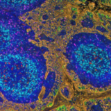
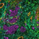
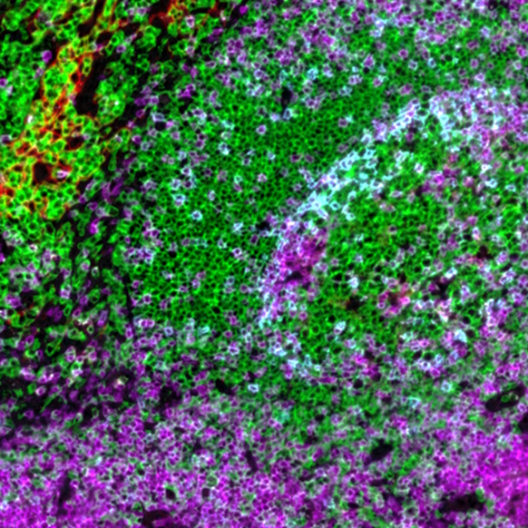
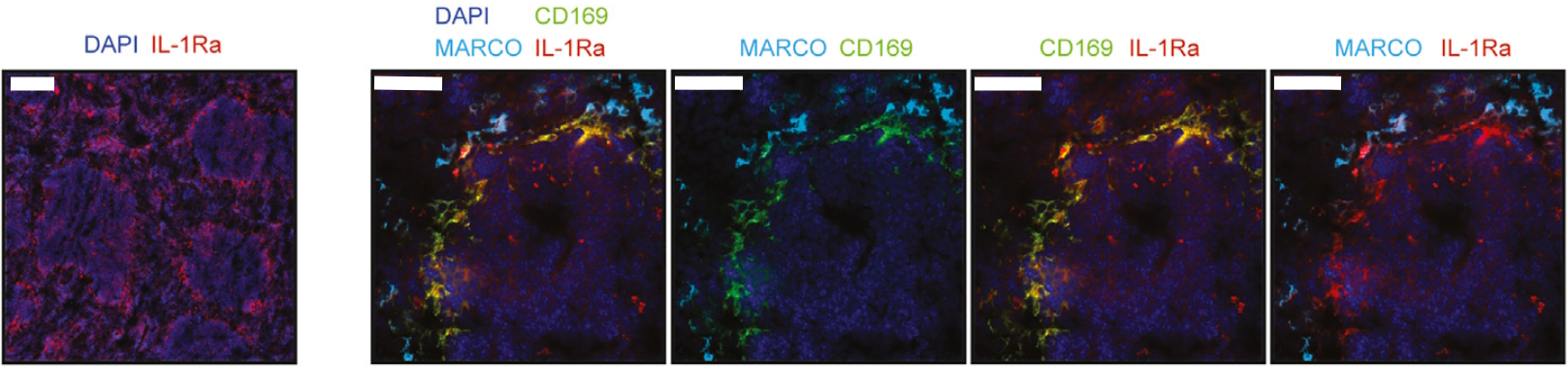
Recently, a research paper published by Gander-Bui et al. facilitated by the LabSat® platform has expanded our understanding of Candida albicans‘ infectious cycle, offering new therapeutic routes to limit its burden2. Authors demonstrated that the interleukin-1 receptor antagonist (IL-1Ra) secreted by macrophages served as a natural immune checkpoint, hindering the effective clearance of pathogens. The IL-1Ra protein is secreted into the bloodstream by CD169+ macrophages in the splenic marginal zone. Conversely, MARCO+ macrophages were observed to lack IL-1Ra expression (Figure 1). Notably, the focused elimination of this molecule resulted in enhanced protection against fatal Candida albicans sepsis. 

Figure 1. Sequential immunofluorescence (seqIF™) on LabSat® determines that IL-1Ra is secreted into the bloodstream by CD169+ macrophages. Scale bars, 200 μm (leftmost picture) and 50 μm (other four pictures). CC License.
Introducing sequential immunofluorescence (seqIF™): a breakthrough in multiplex protein detection 

The study by Gander-Bui et al. used sequential immunofluorescence (seqIF™) on LabSat® to identify CD169+ macrophages of the splenic marginal zone as main producers of IL-1Ra. SeqIF™ presents a novel and straightforward method to detect numerous proteins in a single tissue section with unparalleled efficiency.

Figure 2. Illustration of the workflow of a seqIF™ assay carried out on LabSat®. Once the manual tissue pre-processing is performed, the slide can be loaded into the platform. The system then automates the steps of antigen retrieval and staining cycles. When imaging steps are required, the user takes out the slide, mounts, and images it. If further staining on the same slide must be performed, the user unmounts it and reloads it on LabSat®. The elution step is then automatically followed by a new staining cycle.
What are the main advantages of seqIF™? 
  • Timesaving: seqIF™ allows quick multiplex marker optimization and assay development.
  • Enhanced flexibility: seqIF™ can be performed on FFPE (Formalin-Fixed Paraffin-Embedded) and on FS (Frozen Sections) with customized protocols. Antibodies from the same host species can be used during distinct cycles.
  • Increased multidimensional insights: the most comprehensive data out of precious samples, facilitating the visualization of multiple antigens simultaneously in cellular microenvironments. 

As this method is readily scalable, seqIF™ can be particularly useful in designing immunophenotyping panels where testing of multiple antibodies is required. It allows users to visualize protein co-expression on immune cell subsets to investigate their spatial correlations with functional significance. 

Automate your multiplex assays on LabSat® 

If you are interested in automating manual IHC and IF protocols or if you have other questions related to LabSat® performance in your laboratory, please contact us to determine the best fit for your experimental needs.

References: 
  1. Pappas P. et al. Invasive candidiasis. Nature Reviews Disease Primers. 2018. 4:18026. doi: 10.1038/nrdp.2018.26.
  2. Gander-Bui H. et al. Targeted removal of macrophage-secreted interleukin-1 receptor antagonist protects against lethal Candida albicans sepsis. Immunity. 2023. S1074-7613(23)00280-7. doi: 10.1016/j.immuni.2023.06.023. 

Related Articles