COMET™
Scalable hyperplexing
Unmatched hyperplex throughput with walk-away automation
- Perform a 20-plex on cohorts of 20 samples in just 1 week.
- Virtually unlimited plex level capability (perform multiple additional runs on the same slide).
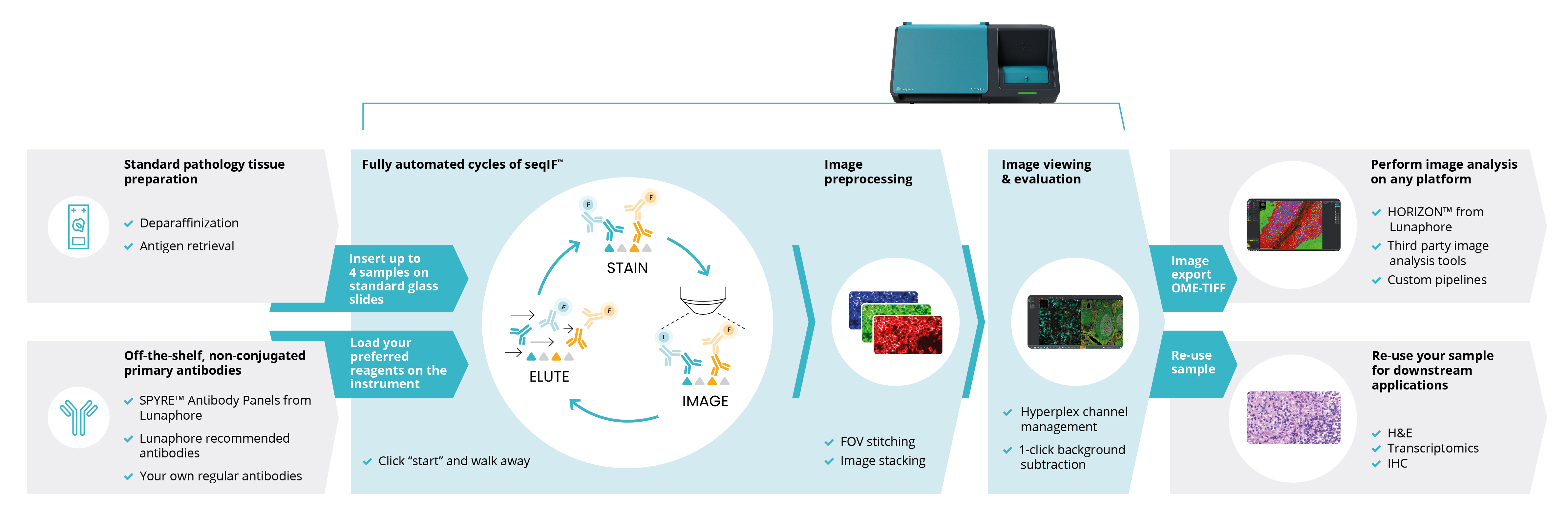
- Slide in, OME-TIFF image out (with background already subtracted).
Rapid and flexible panel development using label-free antibodies
- Use standard, off-the-shelf, label-free primary antibodies. No conjugation or barcoding needed.
- Transfer your existing know-how of IHC / IF antibodies to your COMET™ library.
- Generate hyperplex protocols automatically in just a few clicks.
True reproducibility and tissue preservation
- Maximize reproducibility thanks to a fully automated workflow and precision microfluidics.
- Tissue morphology and epitope stability are fully preserved for downstream applications.
- Avoid undesired variability thanks to no upstream antibody conjugations.
Hyperplex workflow without user intervention
A fully integrated system across staining, image acquisition and image pre-processing.
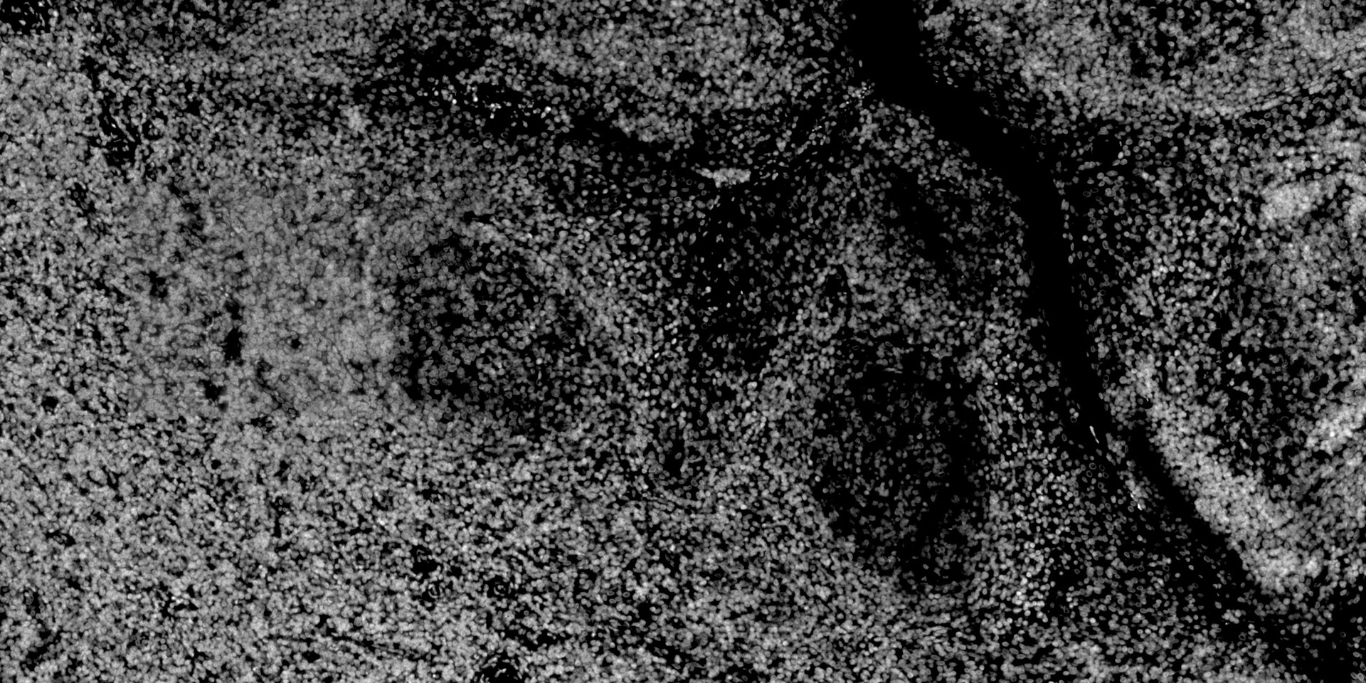
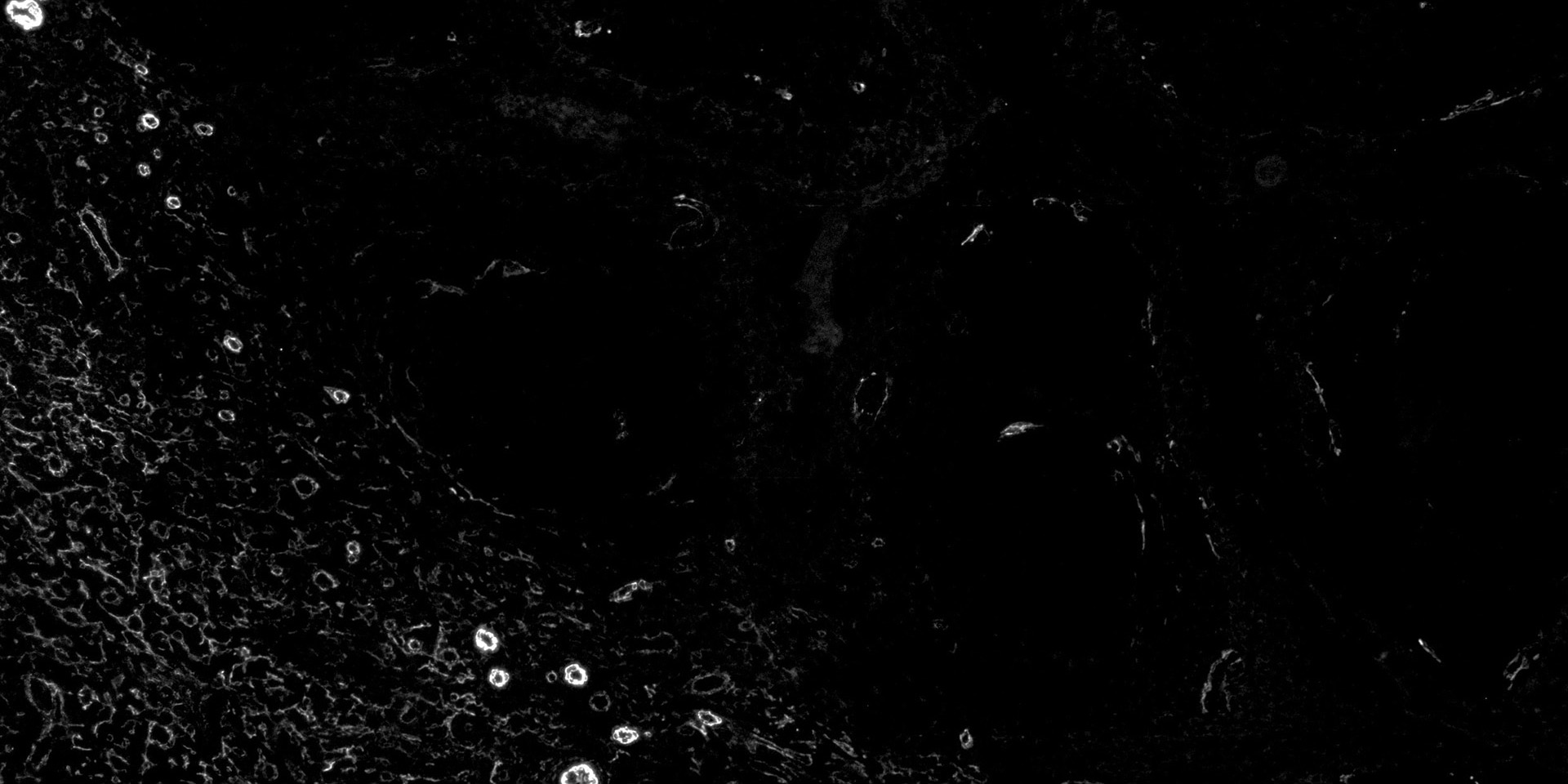
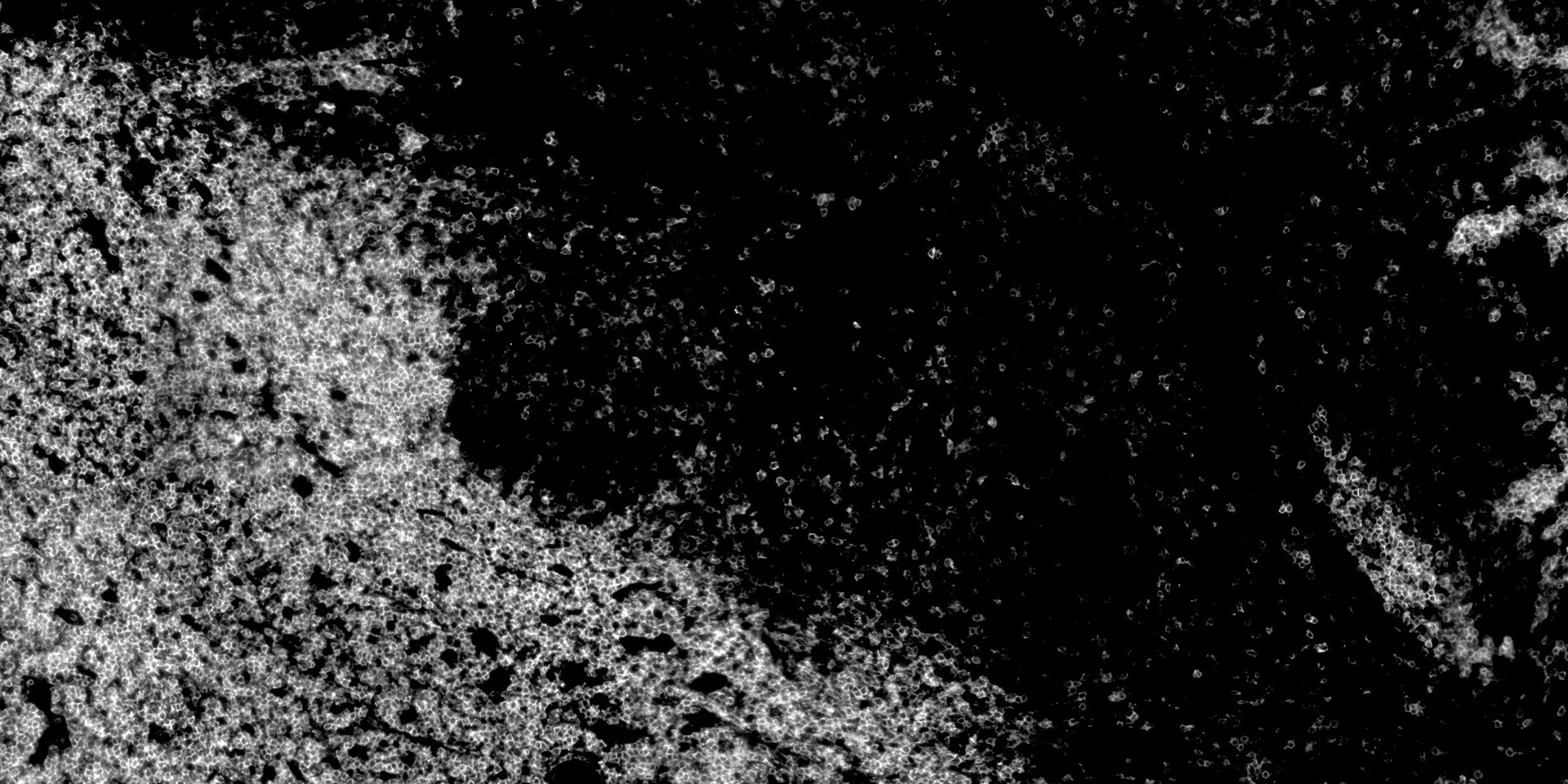
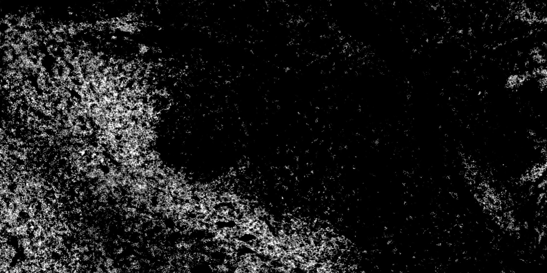
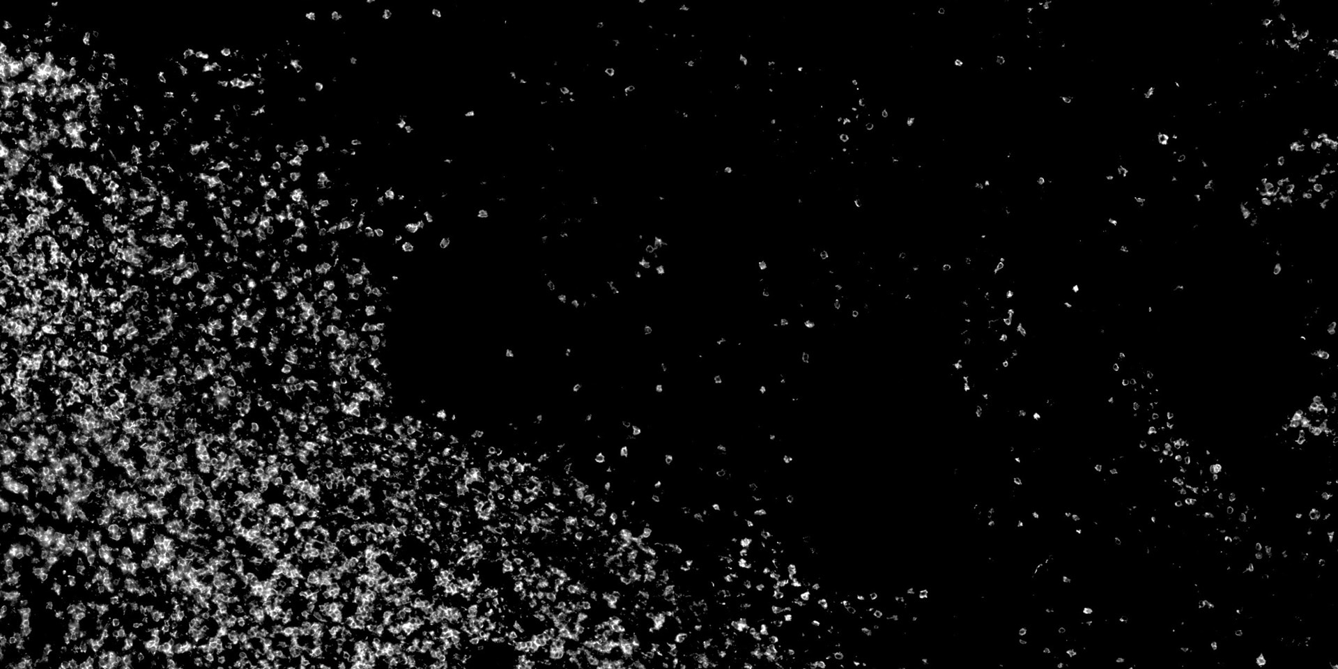
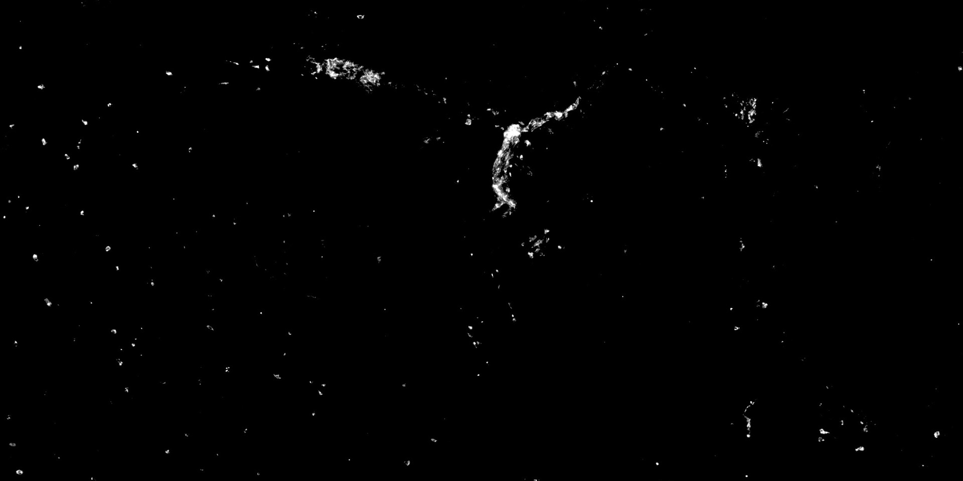
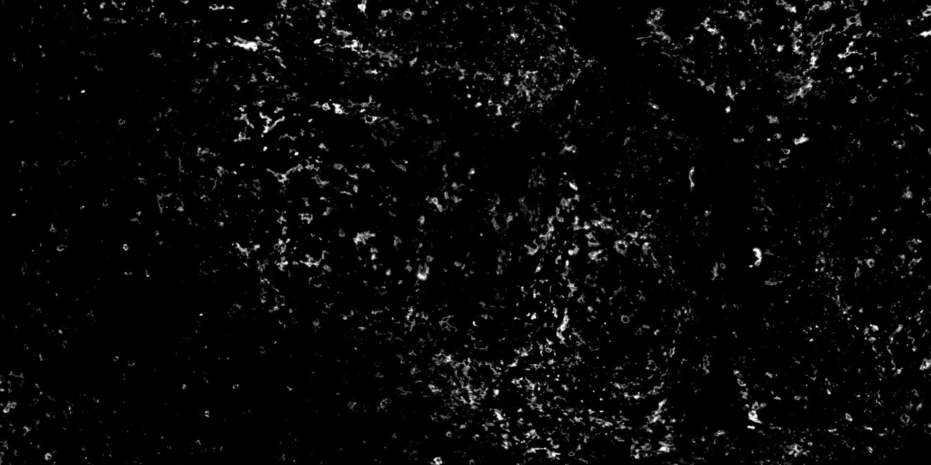
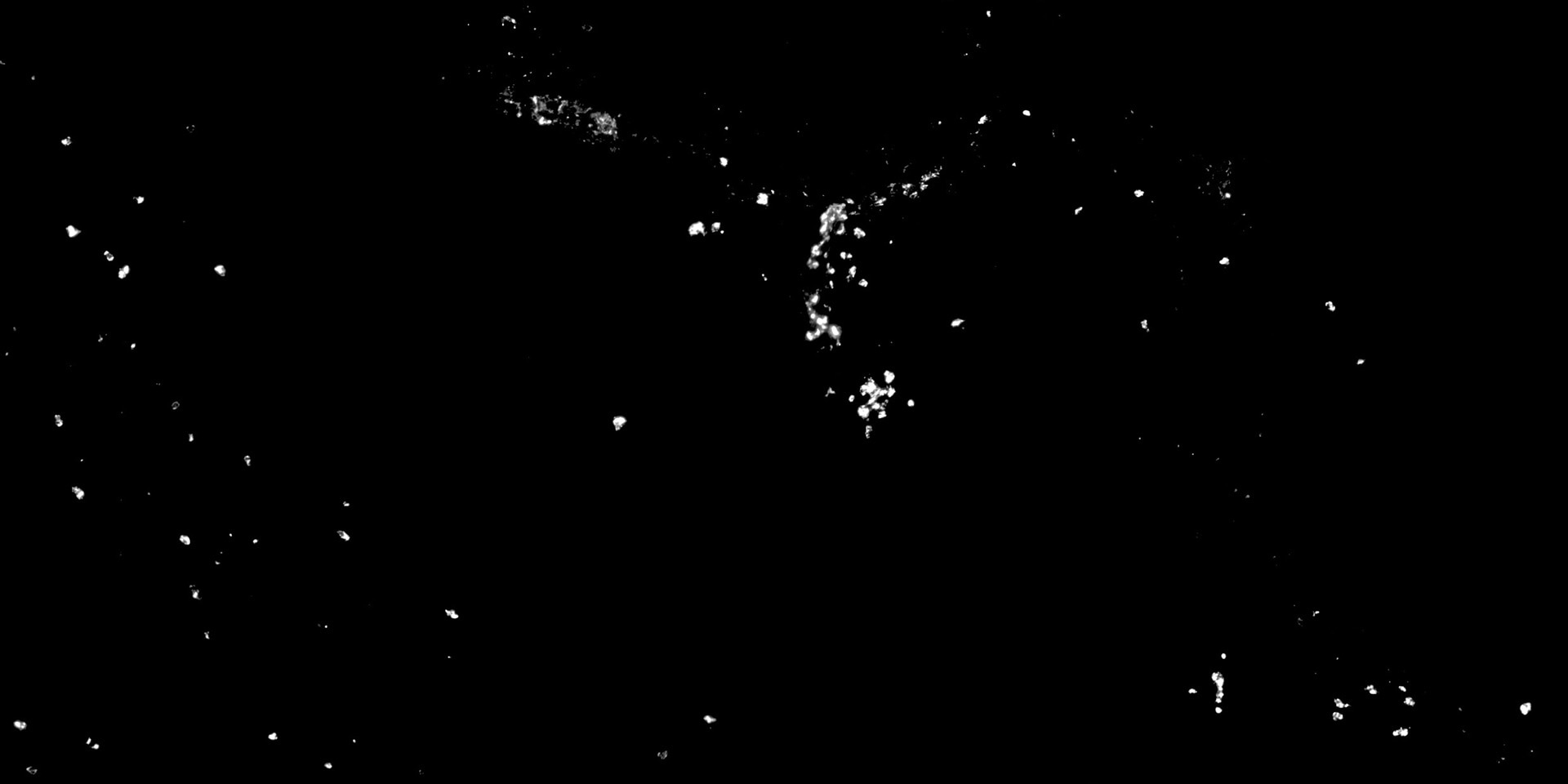
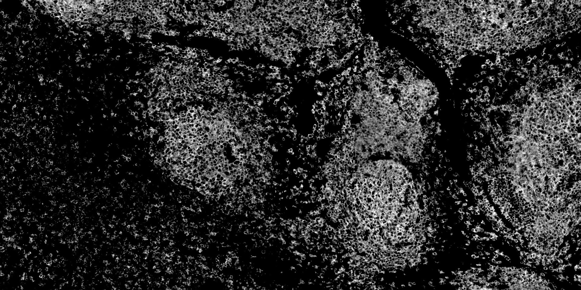
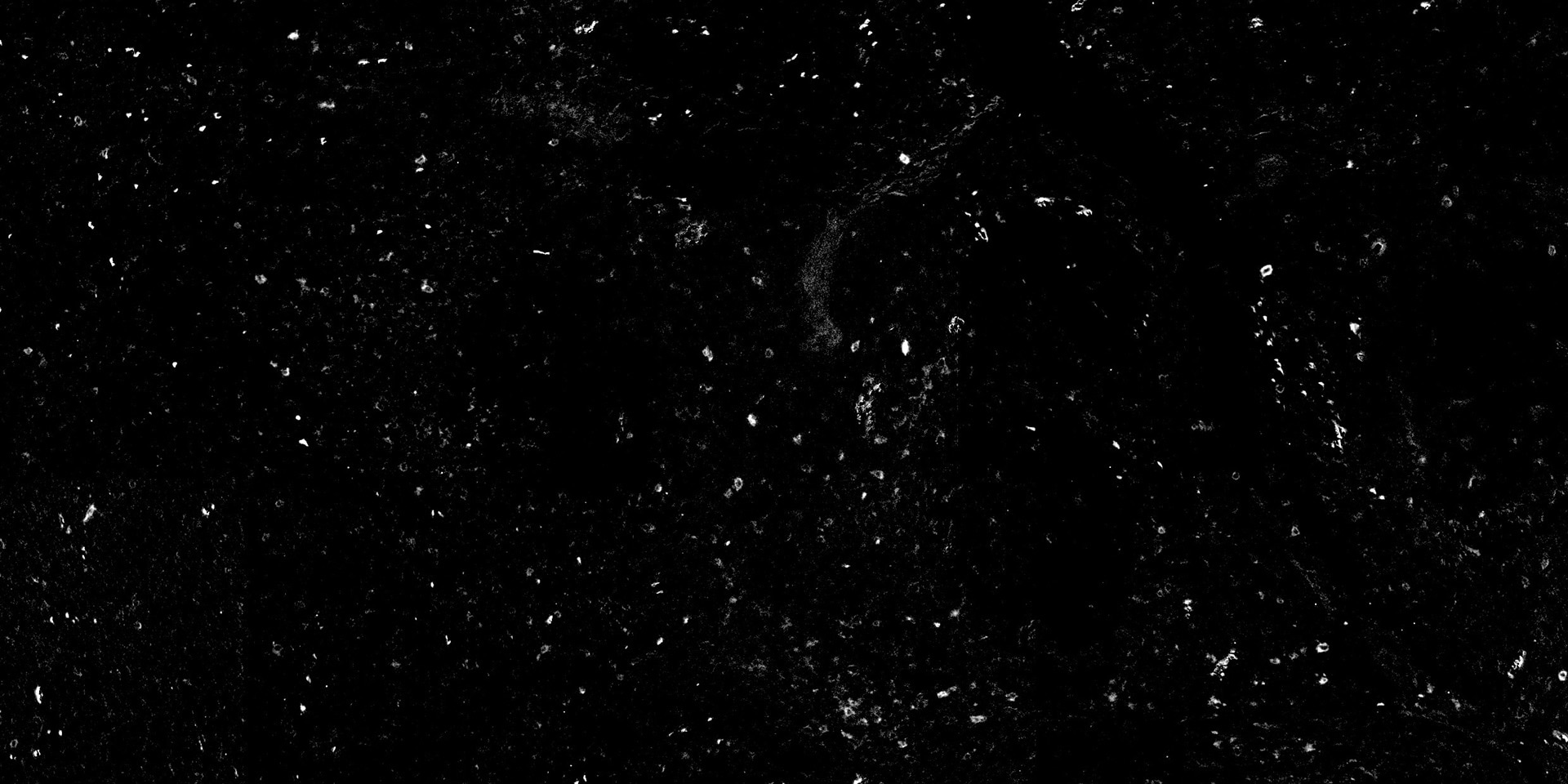
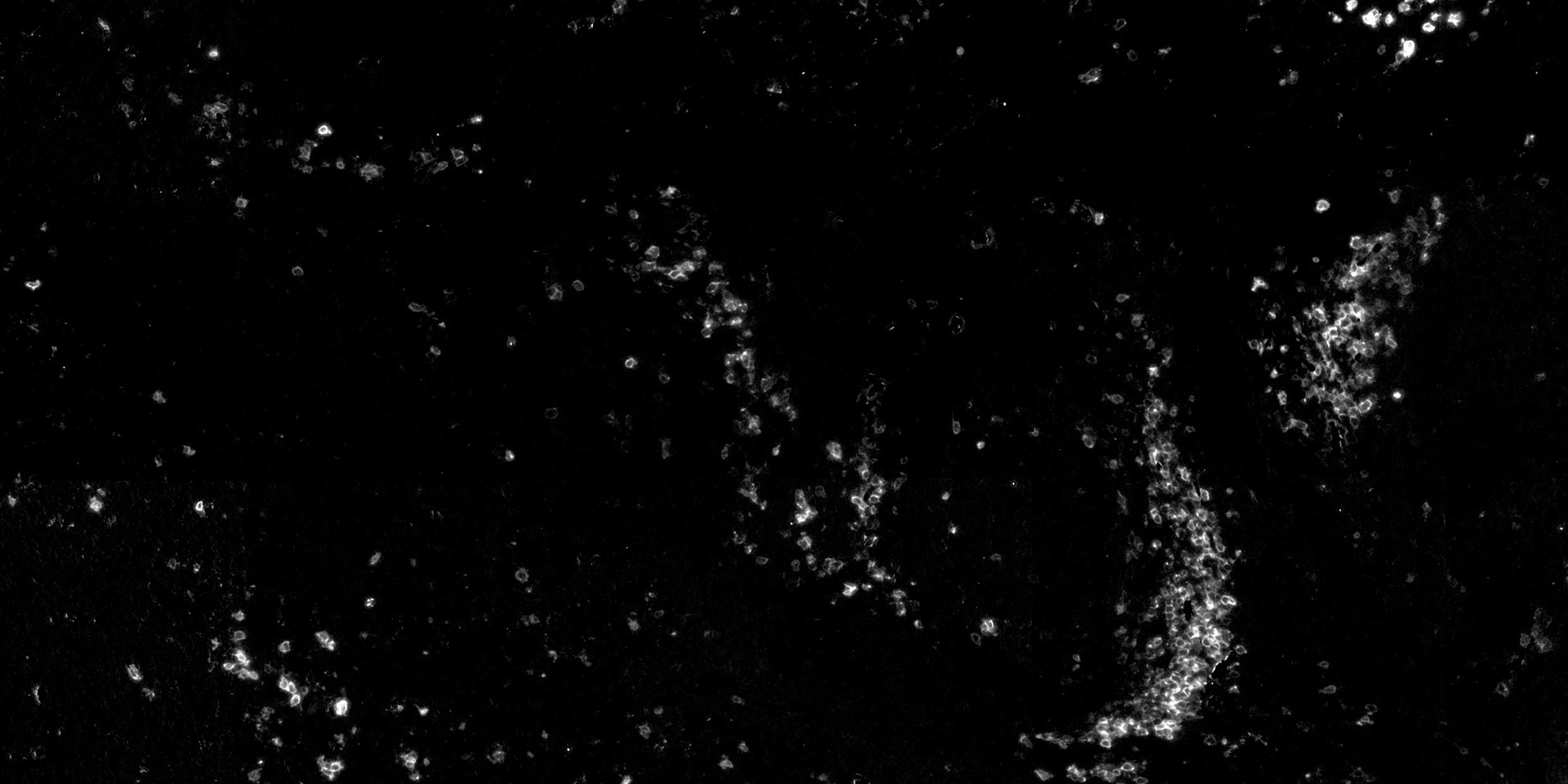
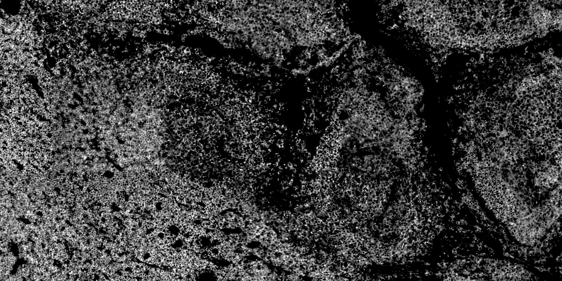
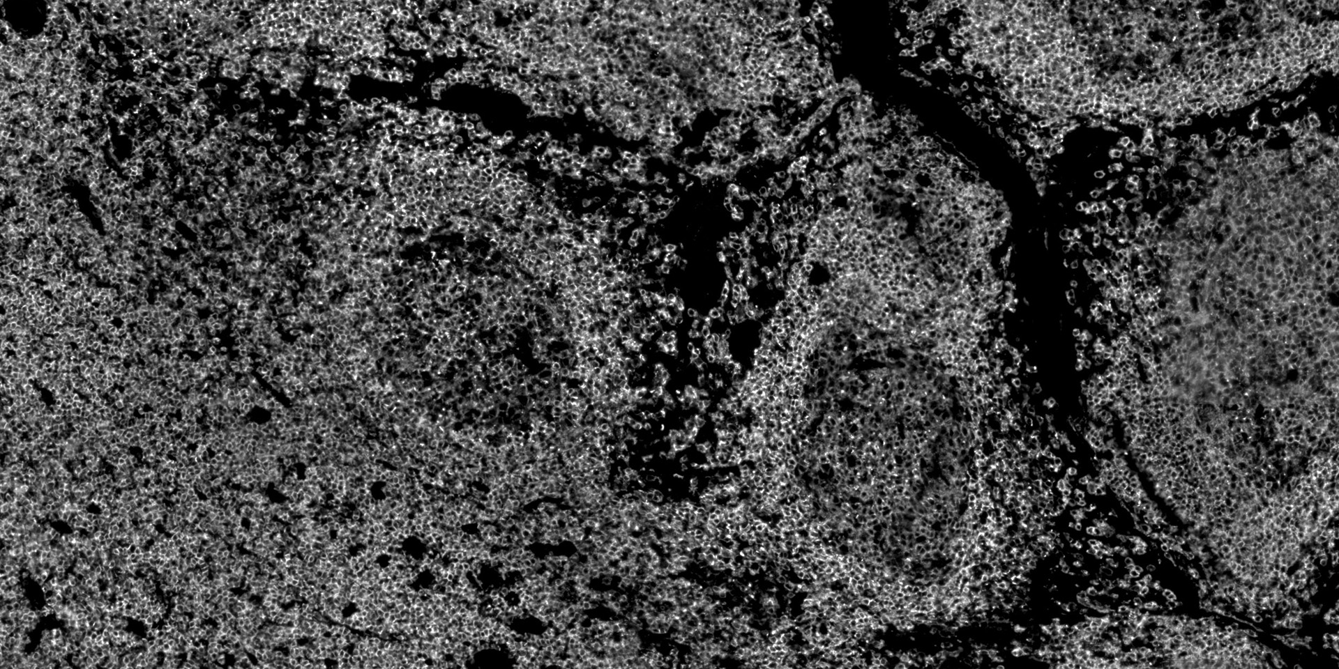
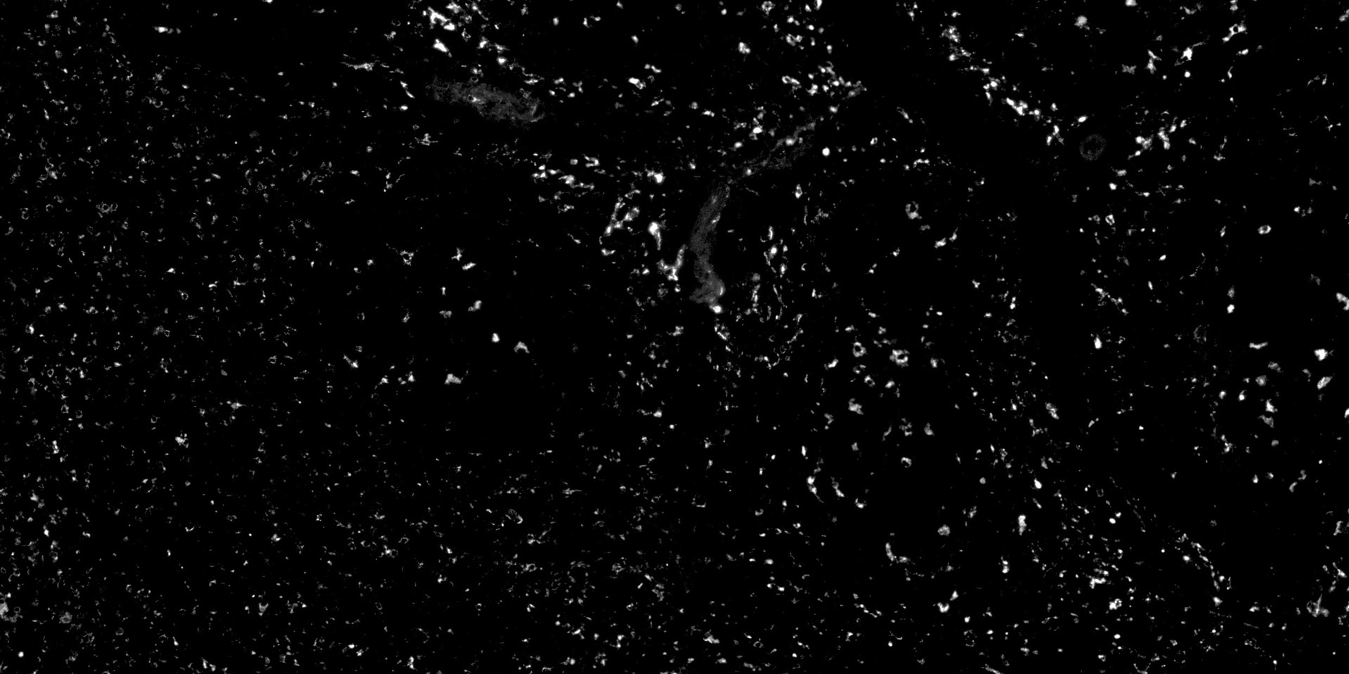
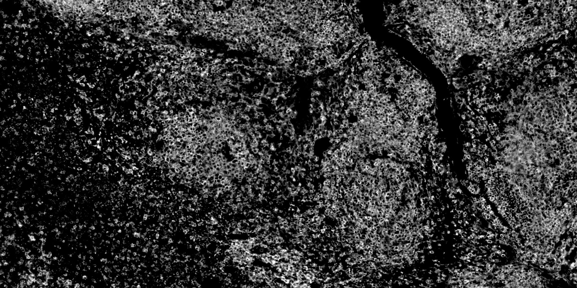
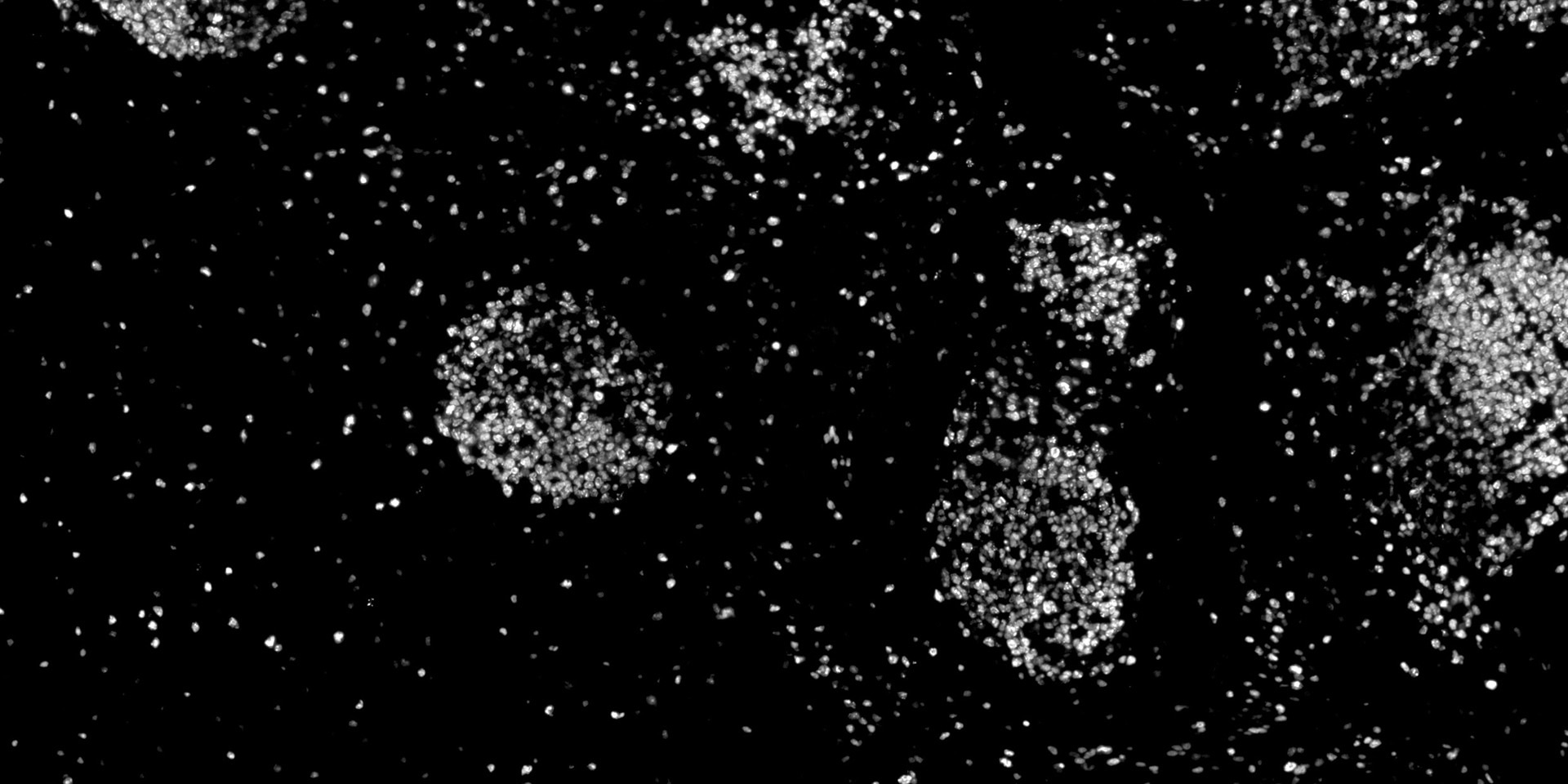
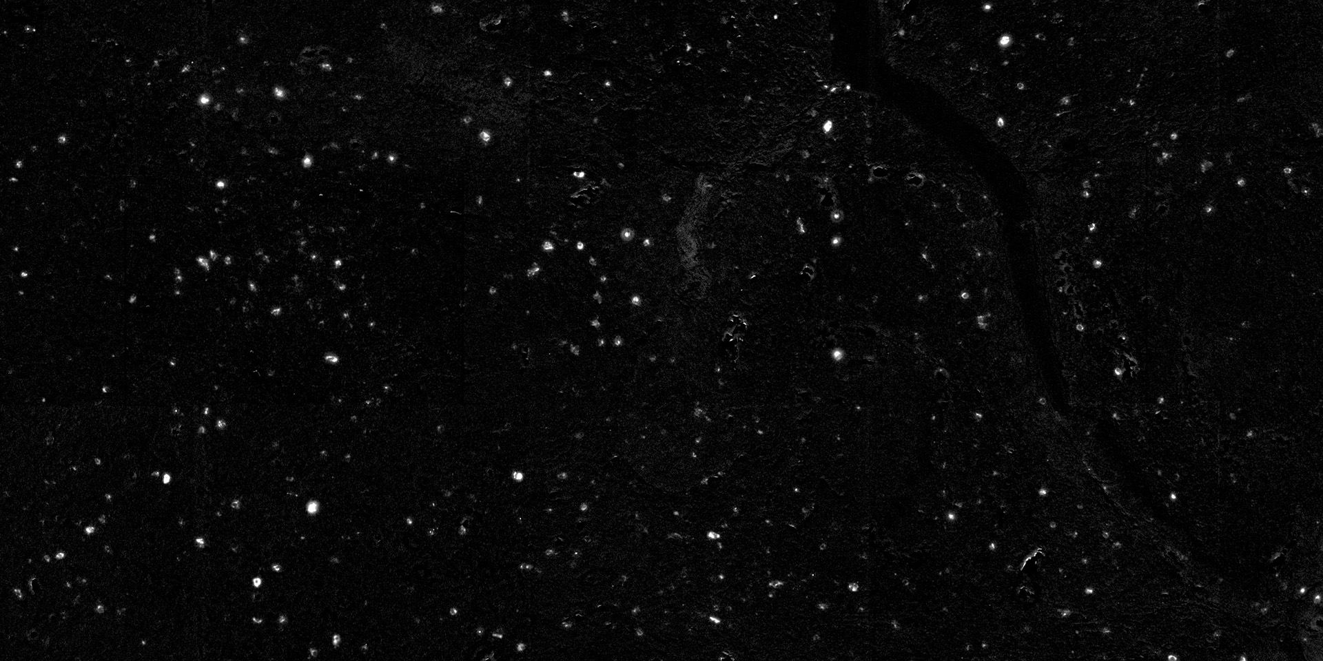
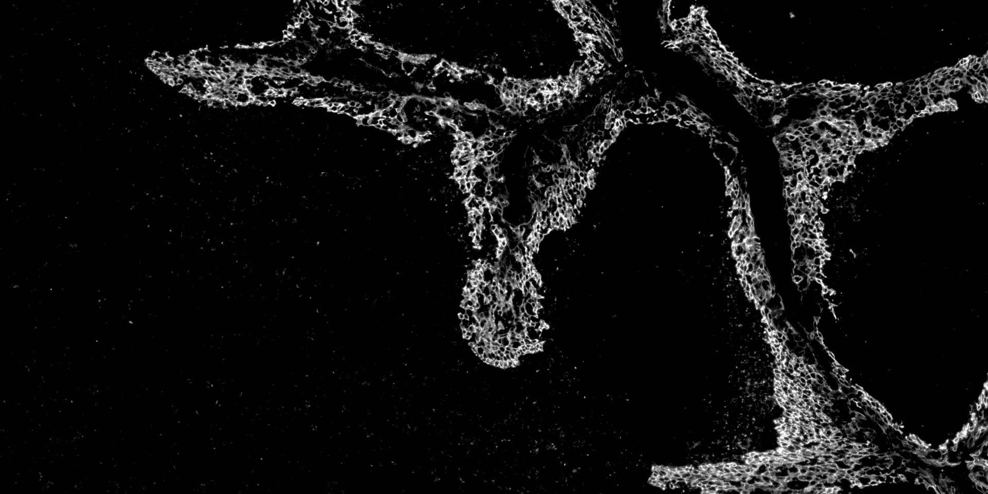
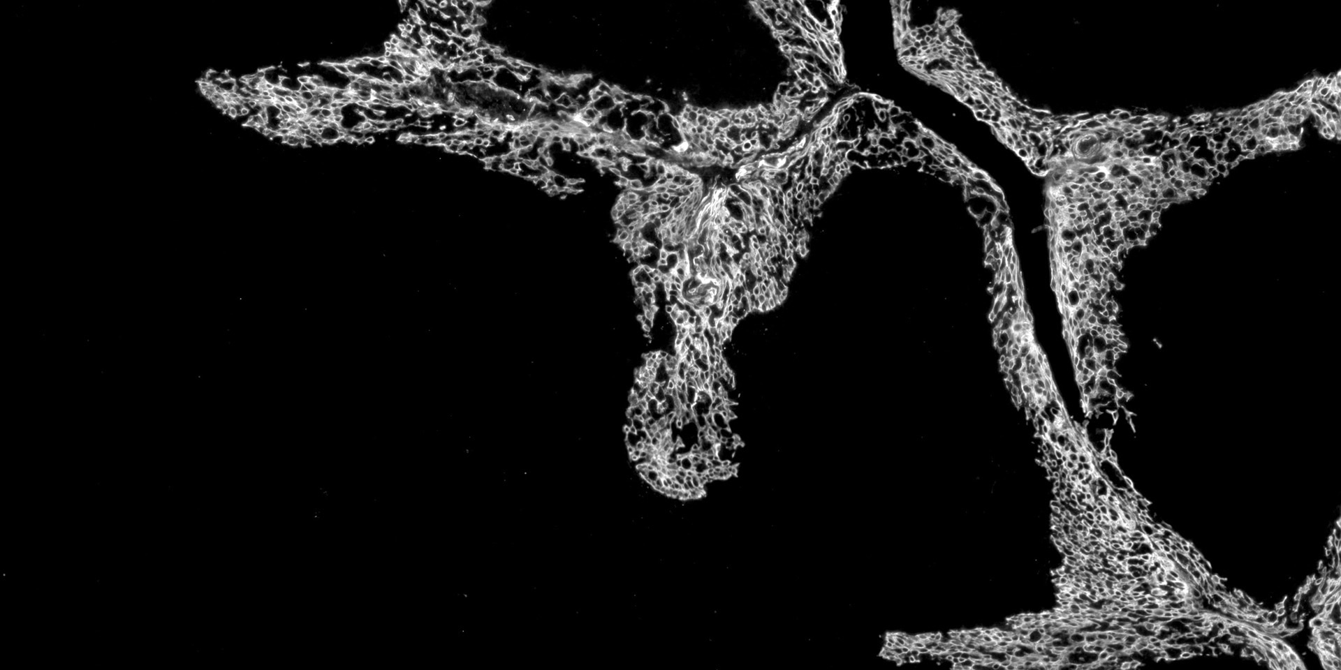
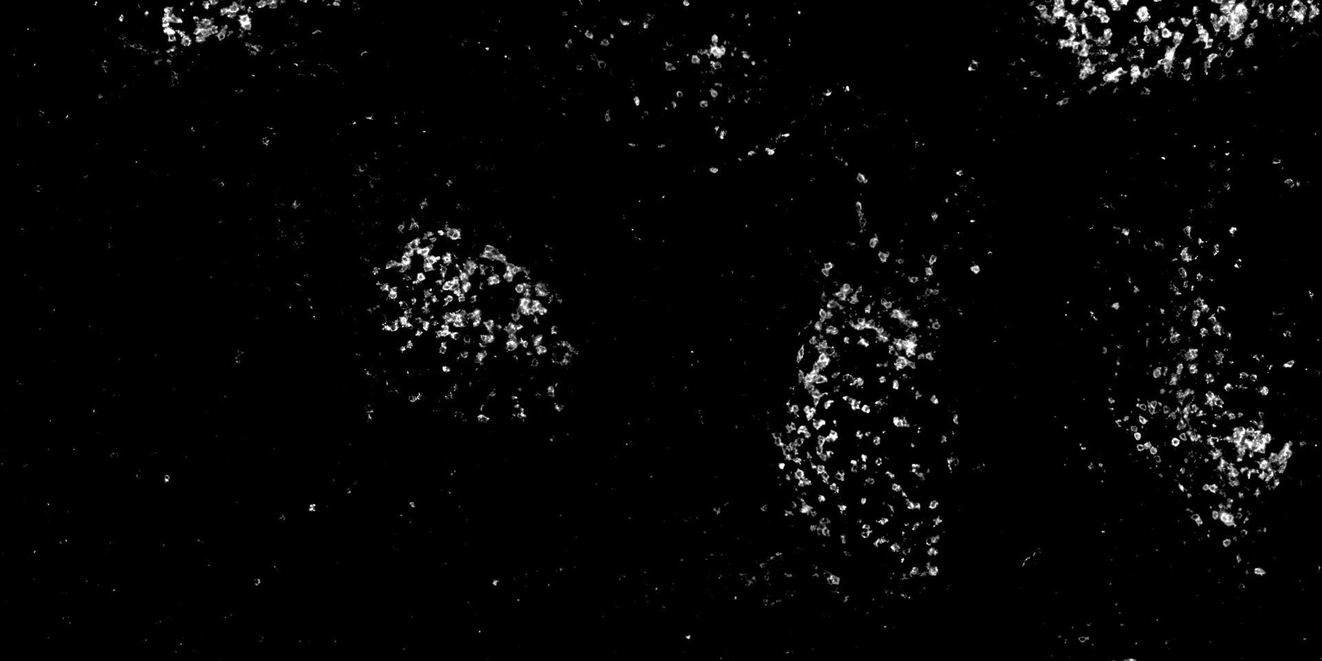
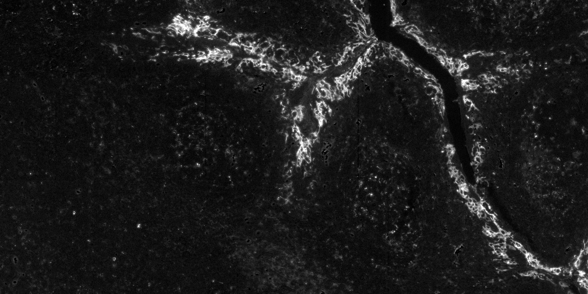
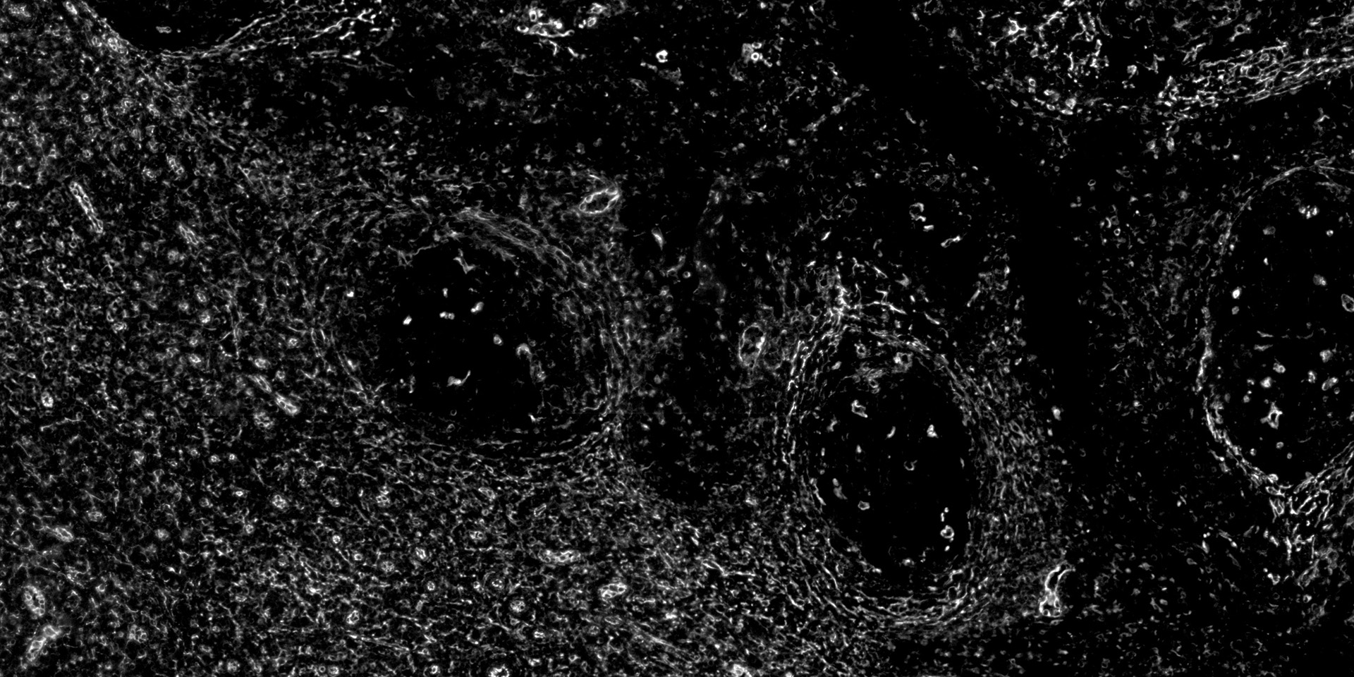
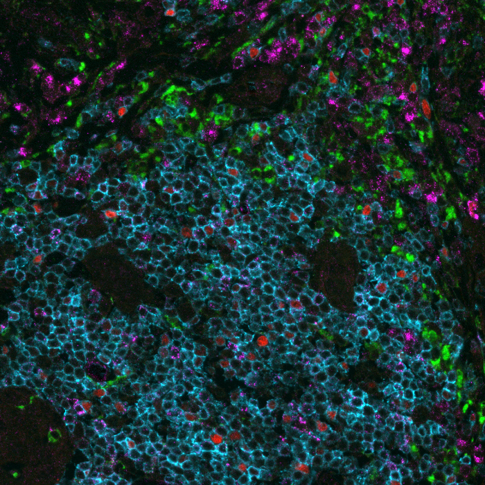
The Lunaphore COMET™ system has enabled us to perform highly-multiplexed imaging of entire cross sections of mouse hearts with sub-cellular resolution. The flexible panel design accelerated antibody validation and measurements so that we can already perform spatial analysis of the heart at scale.
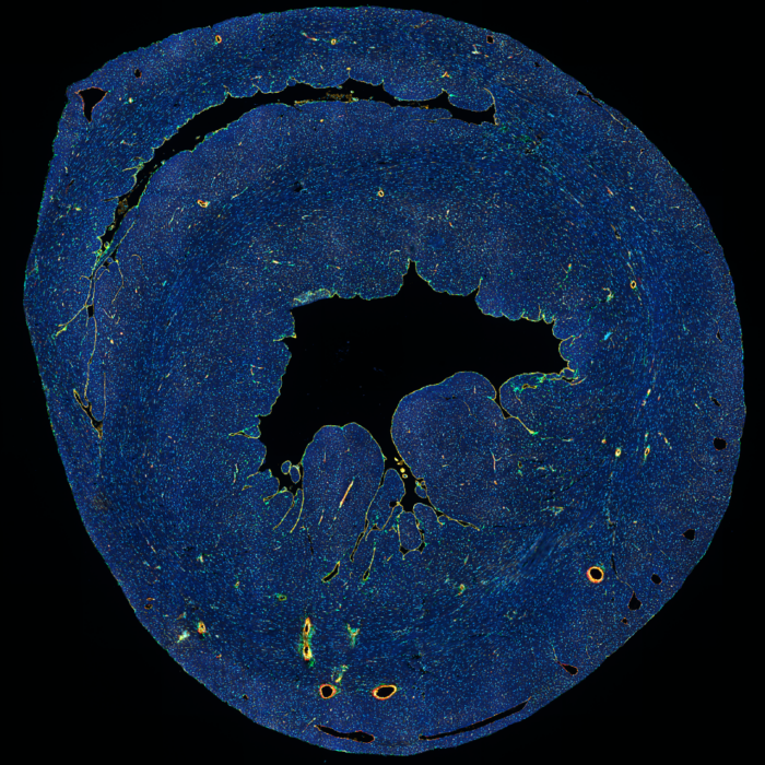
DAPI αSMA CD31 PDGFRa Tnnt2 multiplex image of heart (mouse frozen section)
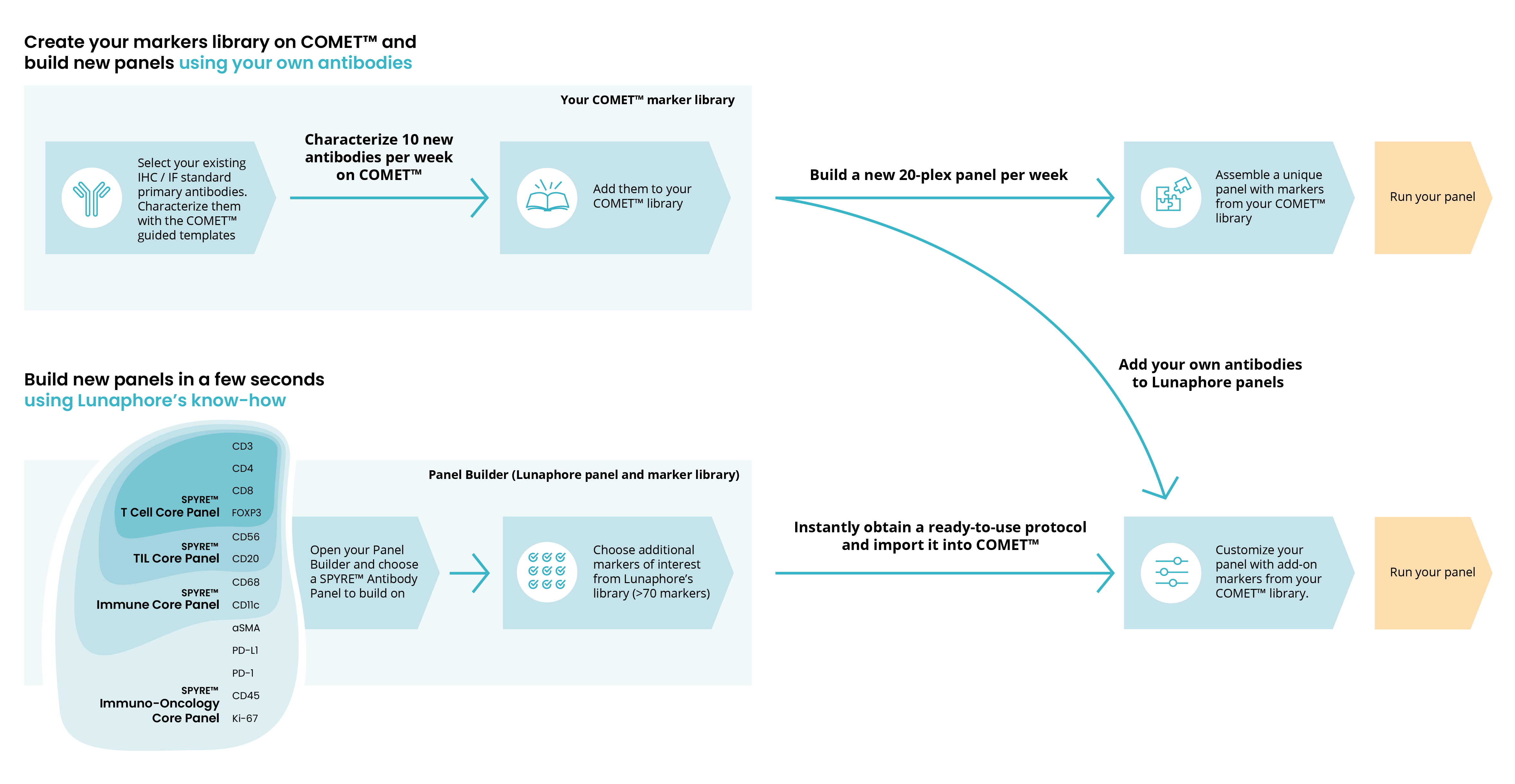
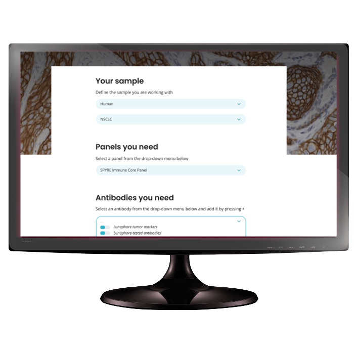
Build custom panels with full flexibility in just a few clicks
COMET™ is:
The first universal, end-to-end, spatial biology solution
A tool to answer your research needs from early discovery to late-stage translational and clinical research. Discover new biological pathways and identify biomarker “signatures” with clinical relevance to support your development of new diagnostic tools and therapies.
A companion in your spatial biology journey
Kick-start your spatial biology adoption with an intuitive and automated platform, and a comprehensive, one-stop-shop, product suite. Be up and running in days.
Your research and development partner
Identify the location and assess abundance of a high number of proteins on a single tissue section, while obtaining large amounts of contextual information. Deep-dive into complex cell interactions in a wide range of applications in immuno-oncology, immunology, neuroscience and infectious diseases.


Tap to toggle on and off.
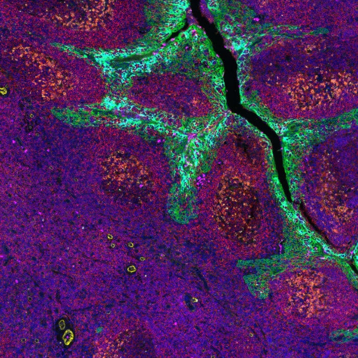
24 markers on tonsil tissue.
Total protocol time: 5h. Fully automated on COMET™.
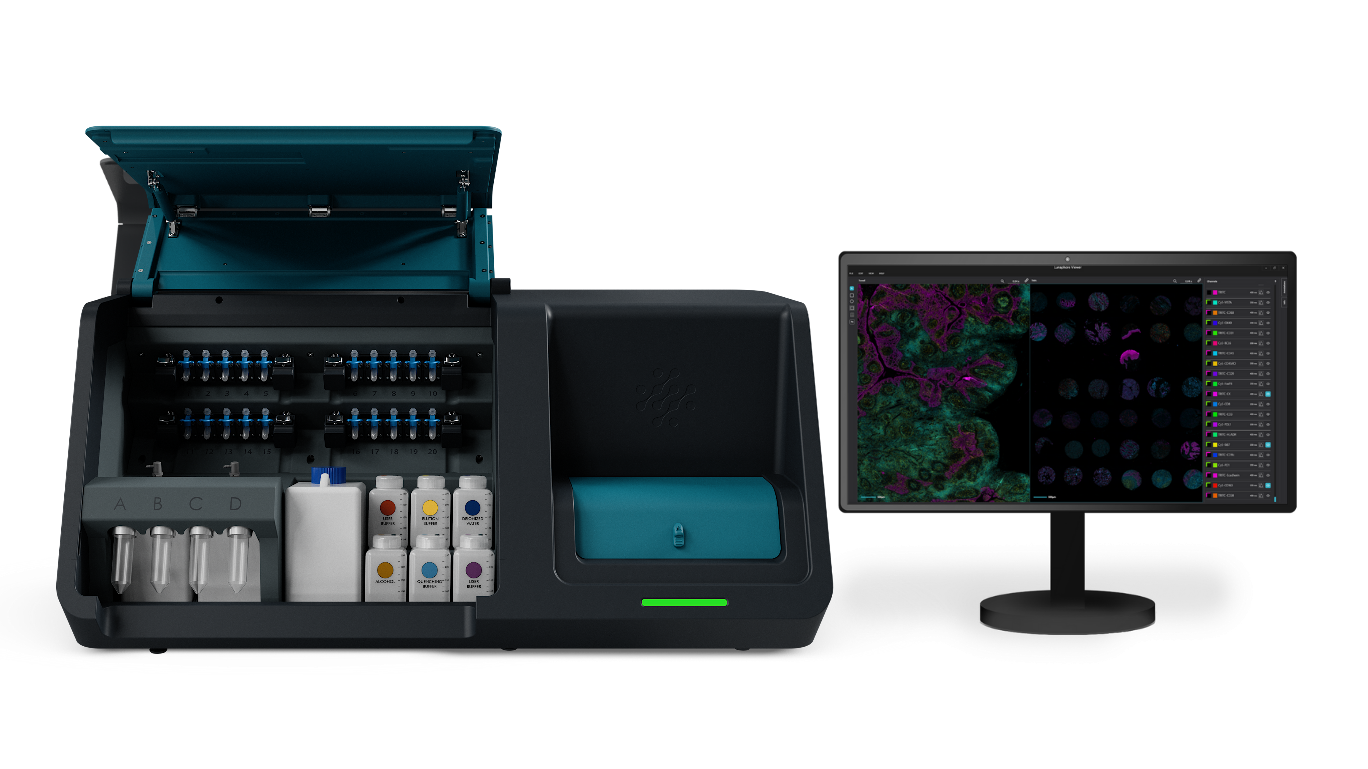
End-to-end: sample in, data out
Click on the buttons to read the description.
-
Reagents Module
Reagents and buffers are loaded in small and large volume reservoirs respectively and delivered to the sample through microfluidic channels.
Close -
Staining Module
Rotary stage where up to four slides are inserted and clamped against the COMET™ Chip. Its unique design allows for staining and imaging parallelization.
Close -
Image Viewer
Following image acquisition, the Viewer can open the resulting image files. The software facilitates visualization and exports the data for image analysis.
Close
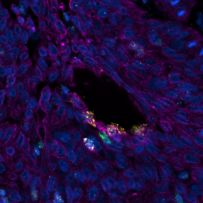
Fully automated multiomics on COMET™
Perform true spatial multiomics assays, simultaneously detecting any RNA and protein targets on the same tissue section using RNAscope™ HiPlex Pro and off-the-shelf, non-conjugated primary antibodies, with subcellular resolution.
A fully automated workflow from target probe hybridization to multiomic imaging, requiring no user intervention, facilitates a deeper understanding of cellular processes and disease mechanisms.
Kick-start your assay development
Run your custom hyperplex panels in days. Kick-start your assay development using the SPYRE™ Antibody Panels:
- T Cell Core Panel
- TIL Core Panel
- Immune Core Panel
- Immuno-Oncology Core Panel
Use the Panel Builder to build ready-to-use protocols in a few clicks, and customize your assays.
Make every marker count
Amplify the signal of low-expressed or hard-to-detect markers with SPYRE™ Signal Amplification Kit. The kit provides the high-sensitivity detection you need without compromising on accuracy.
Analyze your images
Choose your preferred Image Analysis platform for your image.
COMET™ is fully integrated with HORIZON™ by Lunaphore: an entry intuitive tool to start your hyperplex image analysis journey with no coding experience.
COMET™ has proven compatibility with Oncotopix® Discovery (Visiopharm), HALO® & HALO AI™ (Indica Labs), Nucleai AI-powered Solutions and QuPath.
Access Lab
Interested in acquiring a COMET™ solution for your lab?
Simplify your decision making process and technology adoption using your own samples and reagents.