Spatial biology made easy
Multiplex sequential immunofluorescence in a few steps
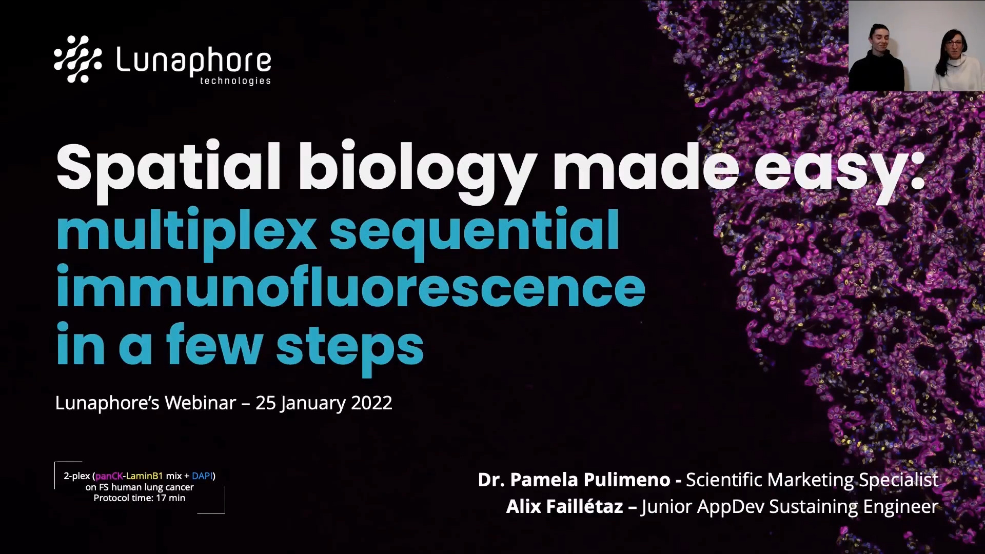
Multiplexing immunofluorescence imaging enables the analysis of the whole tissue microenvironment: the single cell resolution combined with spatial localization retrieves the information of cellular relationships and organization within the same morphological context. However, the complexity of the analysis, time-consuming optimization, execution of staining protocols, and significant reproducibility challenges slow down the wide adoption of such a powerful research tool. In this talk, we introduce how LabSat® performs automated staining and allows to achieve high quality, reproducible, and uniform results thanks to the Fast Fluidic Exchange™ (FFeX™) microfluidic technology. In the second part, our hyperplex platform COMET™ has been adopted to gather comprehensive insights into various spatial biology research applications, such as the biomarker profiling of human prostate cancer tissue by multiplex immunofluorescence assay. Discover how LabSat® and COMET™ can support your research by performing sequential immunofluorescence on the mouse and human tissues from healthy to tumoral samples.
Agenda:
- Multiplexing: why and how;
- Automated multiplexing to accelerate spatial biology research;
- Sequential immunofluorescence on LabSat®;
- Hyperplex immunofluorescence with COMET™ platform;
- Protocol optimization on prostate carcinoma.