Spatial Biology Education
Accelerating Immuno-Oncology Research With A Pre-Configured HORIZON™ Image Analysis Workflow For SPYRE™ Antibody Panels
Posted on:
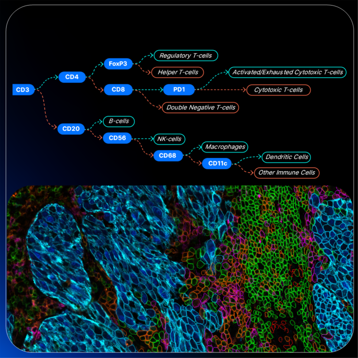
Spatial biology is transforming how researchers study tissues by preserving the spatial context of molecular information. Hyperplex imaging technologies provide an unprecedented view of cellular organization and function by combining protein and RNA detection in the same tissue section.
But while generating this data has become faster and more accessible, extracting meaningful insights remains a significant hurdle.
Hyperplex images are rich in information and are very complex to interpret. Translating them into quantitative, single-cell data requires multiple analytical steps, each with its own challenges. As spatial biology continues to expand, reducing this analytical complexity is key to enable more labs to adopt spatial biology technologies.
COMET™ Suite: A Fully-Integrated Workflow
COMET™ is a high-throughput, multiplex imaging platform that enables spatially resolved, single-cell analysis of protein and RNA expression in tissue samples using sequential immunofluorescence. COMET™ enables researchers to generate high-quality multiomic, hyperplex datasets in a fully automated way.
In parallel, SPYRE™ Antibody Panels simplify assay design by providing modular and ready-to-use antibodies tailored for immuno-oncology applications.
However, the transition from image acquisition to biological interpretation still requires:
- Reliable cell segmentation
- Quantitative feature extraction
- Cell phenotyping
- Spatial analysis
These steps are available in HORIZON™ Image Analysis Software, designed specifically for hyperplex immunofluorescence images generated on COMET™, enabling a smooth transition from tissue staining and imaging to multiplex data interpretation.
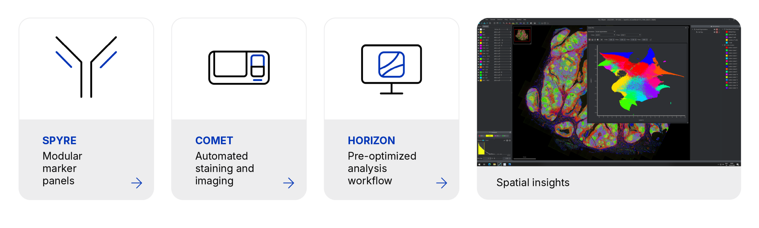
Together, COMET™, SPYRE™ and HORIZON™ create an end-to-end workflow that removes friction at every step from tissue staining to data analysis.
Introducing A Standardized HORIZON™ Workflow for SPYRE™ Antibody Panel Analysis
To support users in navigating the complexity of spatial data, HORIZON™ provides a ready-to-use analysis workflow template optimized for SPYRE™ Antibody Panels for immuno-oncology research. Instead of building pipelines from scratch, users can import the pre-configured workflow and start analyzing their data immediately.
The standardized SPYRE™ analysis workflow integrates the following steps into a single automated pipeline:
- Background Subtraction
Optional background subtraction to improve signal clarity while preserving the integrity of the data. Background channels are automatically selected using metadata available in COMET™ images.
2. Cell Segmentation
HORIZON™ applies COMET™-optimized deep-learning segmentation models to segment nuclei and cells. With the SPYRE™ workflow, the most promising channels for segmentation are pre-selected from available SPYRE™ panel markers.
3. Feature Extraction
Quantitative features are measured across cellular compartments of the tissue sample, enabling detailed characterization of marker expression for downstream cell phenotyping.
4. Feature Thresholding
5. Marker signal intensities are thresholded to separate cells which are positive for a marker from those which are negative. A default threshold value is suggested for markers included in the SPYRE™ Antibody Panel. The default value can be adjusted to better fit the specific staining.
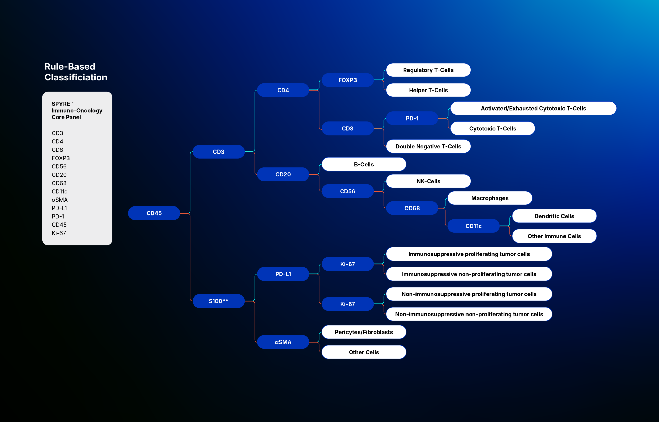
6. Cell Phenotyping
Related Articles