We recently hosted our second annual edition of The Spatial Biology Week™ here at Lunaphore. In live talks, roundtables, and Q&As with key academic and industry experts, we explored the latest advancements in using spatial data in biology, how researchers are overcoming technical challenges, and how world-class innovators are using spatial biology to advance discovery and development of targeted treatments across a variety of diseases.
In our 3-part blog series, we discuss the most important learnings from The Spatial Biology Week™ 2022. As all speakers shared equally essential information to scientific advancement, the insights are presented in no particular order.
The Biological Big Bang
Spatial biology can be compared to the Cambrian explosion – namely, the Biological Big Bang. Before the Cambrian explosion occurred around 540 million years ago, most organisms were relatively simple, composed of single cells, and only occasionally organized into larger groups. After, there was an explosion of different kinds of complex living things that did not resemble anything that had been seen before, many that we can trace to living organisms today.
Dr. Carlo Bifulco, Member and Director of Translational Molecular Pathology and Molecular Genomics at Earle A. Chiles Research Institute, Providence, gave a presentation on Day 1 in which he likened the dramatic appearance of diverse and complex organisms after the Biological Big Bang to the rapid growth of platforms and technology that we are living through today. This is something completely unprecedented that brings us beyond traditional approaches. Where previous methods gave simple readouts – the presence or absence of a biomarker – spatial biology unveils the distribution and neighborhood of a biomarker and how it interacts with the elements around it. The applications, with immuno-oncology (IO), currently being used in spatial biology create the perfect setting for the technology to thrive; as Dr. Bifulco said; “spatial biology has the potential for making a difference in the future and really is already making a difference in the present.”
Spatial biology: “not just a pretty picture”
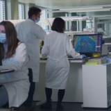
On Day 2 of The Spatial Biology Week™ 2022, we heard about the importance of the image quality that spatial biology technology provides as well as the critical information the image contains – a quantitative dataset.
Kelly Hunter, Chief Scientific Officer at ProPath UK, said, “The images are beautiful, and it’s easy to get caught up in them, but actually, the images aren’t the reason for the experiment. It’s counting the cells, it’s phenotyping the cells, it’s characterizing what they’re doing and where they are, and having something that is analyzable is really important but also excessively analyzable. Not very long ago, people were left with images and left to deal with them in their own time. Platforms being able to support that analysis, particularly by integrating AI tools for segmentation and other things, really make a big difference between getting your results and not just getting a pretty picture.”
Dr. Hunter’s words reinforce just how critical the analytical portion of a spatial biology platform is to speed up our processing capabilities, minimize user error, and provide an unprecedented amount of information to researchers that help move their important work forward.
Standardization creates scientific progress
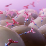
Complex data require powerful solutions. A tremendous amount of information can be retrieved from each spatial immunofluorescence (IF) image, and researchers are working on simplifying and standardizing this process – in many cases, it is not easy.
According to Dr. David Mason, Senior Technical Specialist at Visiopharm, who presented on Day 5 of The Spatial Biology Week™, “high- and ultra-plex data are becoming more common, but image handling and analysis often present some very specific challenges,” such as unsatisfactory cell detection, time-consuming image analysis, lack of confidence in the results, or workflows that require multiple platforms.
To address these challenges, standardization of IF image acquisition and analysis is crucial – including rules for performing cell phenotyping, segmenting cell populations, and analyzing cell neighborhoods. Spatial biology platforms allow for this kind of standardization in the protocol, with pre-built processes that allow for robust and repeatable learning. With these tools, we enable the many different researchers out there to speak the same language and ensure images are captured in a consistent way, facilitating scientific progress through consistent results.