Spatial Biology Education

Uncovering the mechanisms behind immune evasion in pancreatic cancer with multiplex immunofluorescence

Blog: Uncovering the mechanisms behind immune evasion in pancreatic cancer with multiplex immunofluorescence

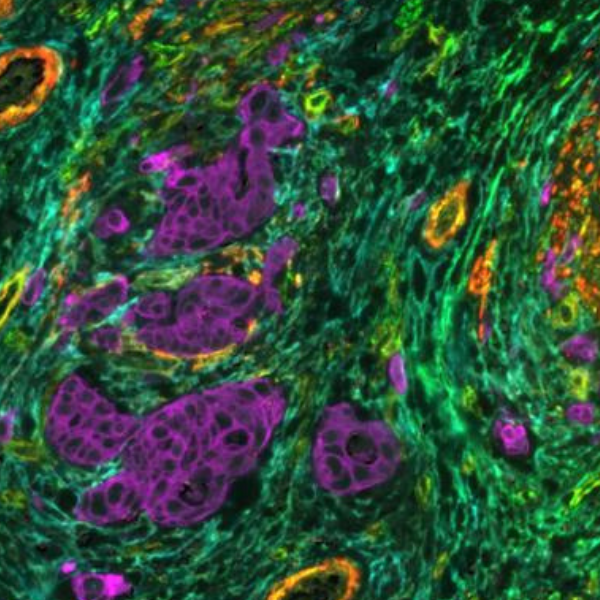
Pancreatic ductal adenocarcinoma (PDAC) is a highly aggressive, fatal cancer1. The typical 5-year survival rate for PDAC tumors is less than 10%, despite scientific advancements in understanding the disease’s biology and the creation of novel therapeutic approaches2. Immune cells play a crucial role in detecting and destroying abnormal cells, but cancer cells can sometimes evade or suppress the immune response. An improved understanding of the tumor immune microenvironment (TME) within PDAC tumors is needed to establish effective immunotherapy treatments. It is known for its highly desmoplastic stroma, an increased growth of connective tissue (or stroma) that happens as a response of the host tissue to invasive cancer cells. Desmoplastic stroma plays a critical role in supporting tumor growth and progression and is thought to contribute to the resistance of PDAC to various therapies. Spatial biology has shown great promise in providing biological insights about this process.

Multiplex immunofluorescence (mIF) as an essential tool in the understanding of PDAC

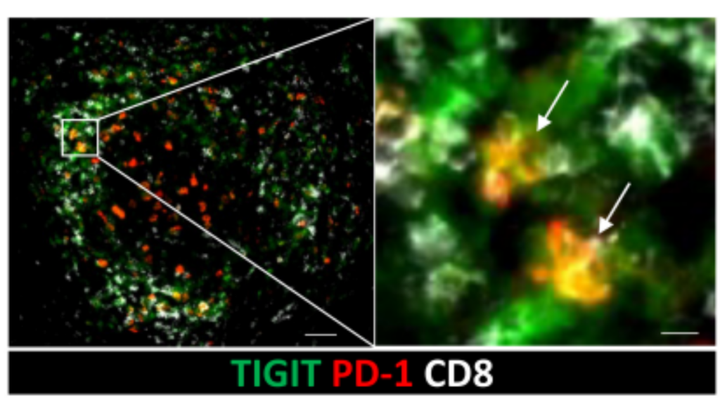
Immune checkpoints are proteins found on the surface of immune cells, such as T cells, and interact with other proteins called ligands, which are found on the surface of target cells, such as some tumor cells. When an immune checkpoint is blocked, it can enhance the immune response, allowing the immune system to attack and eliminate cancer cells. A recent study used mIF, cytometry by time of flight, and single-cell RNA sequencing to characterize tumor-infiltrating T-cell populations in PDAC patients3. Large populations of CD8 expressing tissue-resident memory T cells (TRM) were found. TRMs are a type of T cell that provides a rapid and localized response to antigen challenge4. Antigen challenge refers to the process of exposing the immune system to a specific antigen, which is a molecule that can be recognized by the immune system, such as a protein. These TMRs were expressing PD-1 and TIGIT, two immune checkpoint proteins, at high levels. This discovery was facilitated by performing multiplex immunofluorescence on COMET™ to determine the precise co-localization of two checkpoint proteins from the same TRM cells (Figure 1).

Figure 1. Sequential immunofluorescence (seqIF™) on COMET™ shows the location of PD-1+TIGIT+CD8+ T cells3 (left scale bar: 50 µm, right scale bar: 10 µm). *

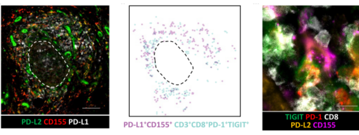
Further authors examined the expression of the ligands for PD-1 (PD-L1 and PD-L2) and TIGIT (CD155 and CD112) on cells within the TME. In ectopic lymphoid structures, PD-L1, PD-L2, and CD155 were expressed in the T-cell region and colocalized with PD-1+TIGIT+CD8+T cells3 (Figure 2).

Figure 2. SeqIF™ on COMET™ of PD-1 and TIGIT ligands within ectopic lymphoid structures (left scale bar: 100 µm, right scale bar: 10 µm). *

PD-1 and TIGIT suppress the activity of T cells, which may contribute to the cancer cells’ ability to evade the immune response. However, the study found that blocking both PD-1 and TIGIT with antibodies can reverse this inhibition and restore the activity of TRM cells. Overall, this research result demonstrates a potential strategy for enhancing the immune response against PDAC consisting in targeting the PD-1 and TIGIT checkpoint proteins on TRM cells3.

Choosing the best technology for your pancreatic cancer research

Multiplex immunofluorescence can be used to study the spatial relationships between different proteins, such as their co-localization within a specific cellular compartment or their distribution within a tissue sample. By analyzing the signals from multiple fluorescent antibody reporters, researchers can obtain contextual information about the location and activity of proteins in the same sample. By leveraging mIF, we can unlock deeper insight into the cell-to-cell variation between and within tumors. This knowledge could then be used to develop diagnostics and treatment strategies in the future of cancer care5.

Technology breakthroughs in the field of spatial proteomics based on an innovative seqIF™ approach on the COMET™ platform, now offer fully automated, quick, and precise multiplex spatial analysis. Integrating COMET™ in a lab allows:

  • fully automated staining, imaging, and image pre-processing to generate highly reproducible data;
  • high-throughput, high-plex spatial analysis, processing 20 slides of 20-plex per week and 40 markers per automated run on the same slide;
  • fast and flexible panel design with automatic panel-building tool using off-the-shelf, label-free primary antibodies;
  • high tissue preservation of samples during analysis, enabling additional downstream modalities such as H&E, IHC, or transcriptomics using the same slide.

COMET™ is compatible with human and mouse samples, Formalin-Fixed Paraffin-Embedded (FFPE), and frozen tissue samples mounted on standard histology glass slides that undergo standard histology pre-processing workflows. With COMET™, the complex cellular interactions and signaling pathways within the TME can be revealed to guide the development of new therapies.

To find out about another interesting study on PDAC do not miss the upcoming webinar from Dr. Julienne Carstens, where she discusses the findings of the tumor regulatory properties of microenvironmental sub-populations and their function-location linkage within the tumor.


References:

  1. Sarantis P. et al. Pancreatic ductal adenocarcinoma: Treatment hurdles, tumor microenvironment and immunotherapy. World J Gastrointest Oncol. 2020. 12(2):173-181. doi:10.4251/wjgo.v12.i2.173.
  2. Miller K. et al. Cancer treatment and survivorship statistics. CA Cancer J Clin. 2016. 66(4):271-89. doi: 10.3322/caac.21349.
  3. Pearce H. et al. Tissue-resident memory T cells in pancreatic ductal adenocarcinoma co-express PD-1 and TIGIT and functional inhibition is reversible by dual antibody blockade. Cancer Immunol Res. 2023. CIR-22-0121. doi: 10.1158/2326-6066.CIR-22-0121.
  4. Szabo P. et al. Location, location, location: Tissue resident memory T cells in mice and humans. Sci Immunol. 2019. 4(34):eaas9673. doi: 10.1126/sciimmunol.aas9673.
  5. Lewis SM, et al. Spatial omics and multiplexed imaging to explore cancer biology. Nat Methods. 2021 Sep;18(9):997-1012. doi: 10.1038/s41592-021-01203-6.


* Work distributed under the Creative Commons Attribution 4.0 International (CC BY 4.0) license.

Related Articles