Poster

Unraveling brain heterogeneity: exploring cellular diversity and molecular patterns with multiplex immunofluorescence

Blog: Unraveling brain heterogeneity: exploring cellular diversity and molecular patterns with multiplex immunofluorescence

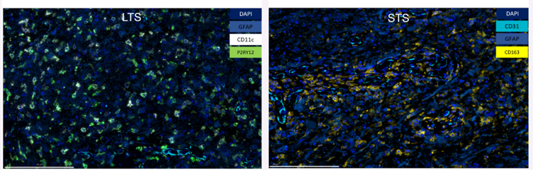
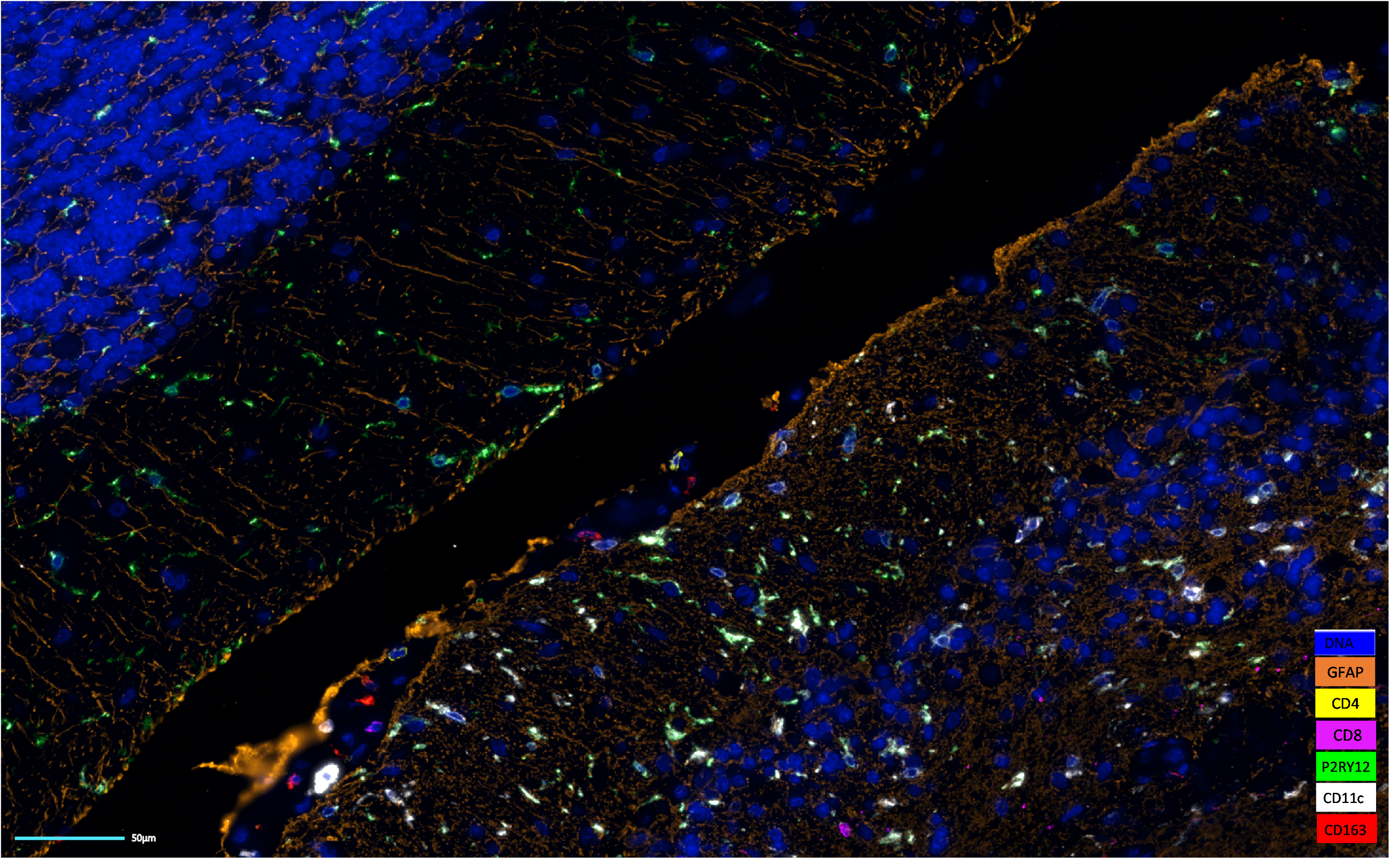
The brain is a complex organ that exhibits cellular and molecular heterogeneity within and between brain regions, especially in a disease setting like a brain tumor. Spatial biology techniques, such as multiplex immunofluorescence (mIF), enable the identification and characterization of cell subpopulations and molecular patterns within the brain (Figure 1). By studying this heterogeneity, researchers can gain a deeper understanding of different cell types’ diverse functions and contributions to brain and disease architecture.

Figure 1. Spatial composition of brain architecture in pilocytic astrocytoma. Image generated on COMET™.
The status of immunotherapy in brain tumors patients

In the context of brain tumors, spatial biology is particularly important because it can provide valuable information about the spatial organization of tumor cells and the surrounding tumor microenvironment (TME). Malignant brain tumors present an array of complex and devastating illnesses, ranging from primary tumors like gliomas to secondary metastases, particularly from non-small-cell lung carcinoma (NSCLC) or breast cancer1. Despite limited therapeutic options, immunotherapy offers a glimmer of hope for individuals battling brain cancer. Although preclinical studies have elicited promising prospects for using immunotherapeutic agents on primary brain tumors like glioblastoma (GBM), clinical trials have yielded ambiguous outcomes. A confluence of complex factors may be attributed to the low efficacy of immune-based therapies in glioblastoma patients. These include genetic and antigenic heterogeneity, reduced mutation loads, and paucity of T cells infiltrating the tumor microenvironment2. However, in contrast to treating primary brain tumors, immunotherapy has proven to be a revolutionary breakthrough in managing brain metastases3. The varied responses to immunotherapies could be attributed to an intricate interplay between molecular and genetic factors influencing T cell antigen recognition and immune responses4.

Immune spatial differences in glioblastoma

In patients with glioblastoma, survival outcomes can vary widely. Studying the immune spatial differences between short-term (STS) and long-term survivors (LTS) can provide insights into the immune response within the tumor microenvironment and potentially identify factors associated with better treatment outcomes. In a recent study presented at Immunology 2023™ AAI Annual Meeting by Dr. Hinda Najem from Northwestern University, researchers profiled TME using sequential immunofluorescence (seqIF™) on COMET™. Authors observed that tumors from LTS are enriched with P2RY12+ microglia, whereas STS are enriched with CD163+ macrophages (Figure 2). Highly activated myeloid cells, expressing either CD8 or CD4, were detected within the TME regardless of survival. Interestingly, in the TME of LTS glioblastoma patients, P2RY12+ microglia interact with naive, non-exhausted T-cells. Therefore, these immune interactions in the TME may help determine STS versus LTS in GBM patients.

Figure 2. mIF on tumors from LTS and STS glioblastoma patients. Tumors from LTS are enriched with P2RY12+ microglia, whereas STS are enriched with CD163+ macrophages.

One of the main challenges in treating brain tumors is their heterogeneity, which means that tumor cells can differ in their genetic makeup, morphology, and behavior. Advances in spatial biology technologies, such as multiplex immunofluorescence, have enabled researchers to study the spatial organization of cells and molecules at high resolution. This has opened new opportunities for understanding the complex biology of brain tumors and identifying more effective treatments.

Download the poster to:

  • Discover how hyperplex on COMET™ allows for reproducible assessment of biomarkers in glioblastoma patients.
  • Find the full list of markers used in the study.
  • Learn about the frequency of immune cells between STS and LTS cohorts.

References:

  1. Quail DF and Joyce JJ. The Microenvironmental Landscape of Brain Tumors. Cancer Cell. 2017. 31(3):326-341.doi: 10.1016/j.ccell.2017.02.009.
  2. Buerki RA et al. Immunotherapy of primary brain tumors: facts and hopes. Clin Cancer Res. 2018. 24(21):5198-5205.doi: 10.1158/1078-0432.CCR-17-2769.
  3. Kluger HK et al. Long-Term Survival of Patients With Melanoma With Active Brain Metastases Treated With Pembrolizumab on a Phase II Trial. J Clin Oncol. 2019. 37(1):52-60. doi: 10.1200/JCO.18.00204.
  4. Friebel E. et al. Single-Cell Mapping of Human Brain Cancer Reveals Tumor-Specific Instruction of Tissue-Invading Leukocytes. Cell. 2020. 181(7):1626-1642.e20. doi: 10.1016/j.cell.2020.04.055.

Related Articles