Infographic

Spatial biology applications: obtain insights into biological processes with spatial proteomics

Blog: Spatial biology applications: obtain insights into biological processes with spatial proteomics

Are you getting started in spatial biology and wondering if this is the right solution for your research? Discover our infographic and learn more.

Spatial biology allows researchers to observe and measure cellular architecture. It facilitates understanding of the cellular function and cell-to-cell interactions. One of the most common subcategories of spatial biology is spatial proteomics. It entails a variety of unique approaches for obtaining information on different cell types and their dynamics.

Spatial biology enables a wide range of applications

Thorough knowledge of biological functions requires an understanding of the spatial context. Therefore, the findings that are made possible by spatial biology have the potential to transform our knowledge in many fields with key areas including:

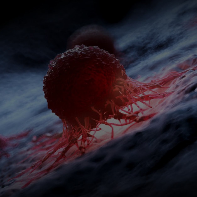
Cancer research: In recent years, cancer treatment has been transformed by immunotherapy and patients have demonstrated encouraging results in several cancer types. The spatial approach allows the identification of specific and predictive biomarkers of response to therapy to match patients to the right treatment. In addition, it facilitates clinical oncologists to identify non-responders earlier and make better decisions.

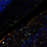
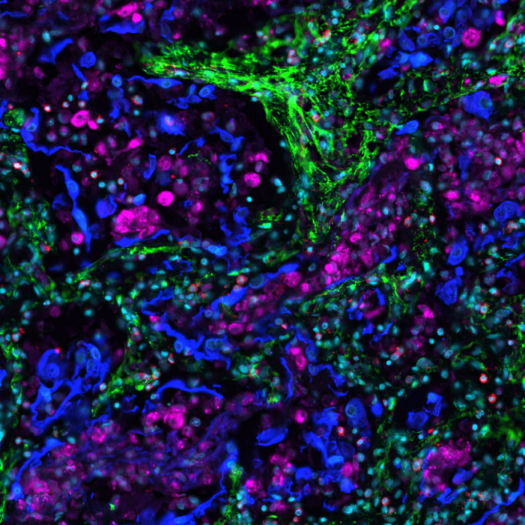
Spatial biology tools also facilitate discoveries of new therapeutic targets in oncology to meet unmet clinical needs. For example, brain tumors are characterized by the immunosuppressive tumor microenvironment (TME)1 and high myeloid cell content2. Therefore, many patients with brain metastasis (BrM) are unable to profit from the development of immunotherapies and must rely on standard-of-care medicines with low success rates. Protein-based spatial biology allows for the characterization of neural tissue in detail. This knowledge will provide insights into therapy resistance and will aid the development of effective medicine.

Neuroscience: Multiplex, high-quality data allow cell-type evaluation in neuroscience and improve our understanding of how the brain works. With this degree of knowledge comes the ability to better understand and treat neurological disorders. For example, in multiple sclerosis (MS), spatial biology tools are allowing us to understand microglia heterogeneity and uncover several molecules that may play a role in pathogenesis3.

Infectious diseases research: Spatial proteomics methods are used to look at pathogen distribution, identify infected and uninfected cell types, aid to understand the phenotype of immune infiltrates in infected tissues, and dissect virus-specific host responses (Figure 1).

Figure 1. Image of the lung tissue in Sars-CoV-2-infected patients with a 24-plex panel. Spatial biology allows researchers to better understand disease mechanisms.

Thanks to spatial biology, researchers can understand that communication within the tissue is essential to comprehend disease causes and its progression and identify the best therapeutic approaches.


Download the infographic to discover:
  • How spatial biology facilitates the study of the tumor microenvironment (TME).
  • Examples of the key applications in protein-based spatial biology, also known as spatial proteomics.
  • Data showing high-quality results from healthy to tumoral samples.
  • Tips for selecting the right tools for spatial proteomics studies, which can facilitate your decision-making process.

References:

  1. Friebel E, et al. Single-Cell Mapping of Human Brain Cancer Reveals Tumor-Specific Instruction of Tissue-Invading Leukocytes. Cell. 2020; 181(7):1626-1642. doi: 10.1016/j.cell.2020.04.055.
  2. Niesel K, et al. The immune suppressive microenvironment affects efficacy of radio-immunotherapy in brain metastasis. EMBO Mol Med. 2021; 13:e13412. doi: 10.15252/emmm.202013412.
  3. Miedema A, et al. High-Resolution Transcriptomic and Proteomic Profiling of Heterogeneity of Brain-Derived Microglia in Multiple Sclerosis. Front. Mol. Neurosci. 2020; 13:583811. doi: 10.3389/fnmol.2020.583811.

Related Articles